
the Creative Commons Attribution 4.0 License.
the Creative Commons Attribution 4.0 License.
 | 05 Oct 2026
Reviews and syntheses: Snow algae on the move – biased motility and snowpack interaction from a biophysics perspective
Caitlin S. de Vries

Melody J. Sandells
Matthew P. Davey
Gary S. Caldwell
Ottavio A. Croze
Snow algae are psychrophilic and psychrotolerant photosynthetic microorganisms found on every continent, predominantly in polar and alpine environments. Along with contributing to terrestrial carbon cycling and food webs, colourful snow algal blooms formed on snow surfaces can substantially reduce albedo and accelerate snowmelt. Despite their ecological importance, the mechanisms governing snow algae motility and migration within snow remain poorly understood. This review synthesises current knowledge of snow algae migration, spanning microscopic cell-level motility to macroscopic population-level redistribution within snowpacks. We consider snow algae as biologically active particles within the framework of active matter physics, exploring their non-equilibrium dynamics and self-propelled motion in response to environmental stimuli. Particular attention is given to directional behaviours in response to light, temperature and chemical gradients, gravity and fluid flow. Where data gaps exist, we draw parallels from studies on a model motile microalga, Chlamydomonas reinhardtii. Finally, we identify key knowledge gaps and highlight future research directions, with implications for understanding cryosphere processes, microswimmer tactic behaviour, and the development of emerging biotechnologies.
- Article
(4491 KB) - Full-text XML
- BibTeX
- EndNote


Snow algae, which give rise to the terms blood snow and watermelon snow, are visually striking and ecologically significant photosynthetic microalgae (Fig. 1). Found on every continent, primarily in polar and alpine environments (Duval et al., 1999; Remias et al., 2005), these keystone primary producers act as terrestrial carbon sinks (Gray et al., 2020), facilitate nutrient cycling (Sommers et al., 2026), influence microbial community dynamics (Brown et al., 2015; Tucker and Brown, 2022), and provide nutrition for higher organisms (Ono et al., 2021; Sugden et al., 2025). Through a reduction in albedo, snow algae can accelerate snow melt rates (Ganey et al., 2017; Khan et al., 2021; Roussel et al., 2024; Thomson et al., 2025), potentially contributing to shifts in local hydrology (Hoham and Remias, 2020). Snow algal proliferation is linked to meltwater presence and the subsequent release of nutrients (Hoham and Ling, 2000; Hoham and Mullet, 1977). How these relationships may respond under future climate change scenarios remains an important question and requires further investigation. Despite snow algae's global significance, the mechanics behind how they actively migrate through complex, porous and evolving snowpacks, their primary habitat, remains poorly understood.
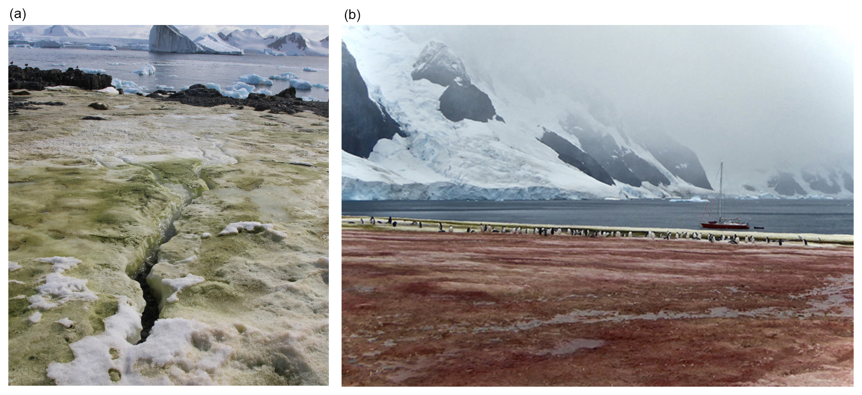
Figure 1Surface snow algal blooms in Antarctica. (a) A surface green algal bloom, Ryder Bay, Antarctic Peninsula. Photo credit: Matthew Davey. (b) Surface red and green snow algal blooms, Antarctic Peninsula. Photo supplied by the National Snow and Ice Data Center, University of Colorado, Boulder. Photo credit: Bob Gilmore.
Partly due to their psychrophilic (optimum temperature range of 0–10 °C) and psychrotolerant (tolerate temperature ranges up to ∼ 20 °C) nature (Gonzalez et al., 2026; Hoham, 1975; Moyer et al., 2017; Suzuki et al., 2023), snow algal communities are attracting attention from the agri- and biotechnology sectors for potential outdoor cultivation in low-temperature climates (Hulatt et al., 2017; Leya et al., 2009; Schoeters et al., 2022), where their growth reduces or eliminates the energy input typically required to maintain optimal temperatures for mesophilic species. Taxa such as Sanguina sp. (Procházková et al., 2019) are rich in the red keto-carotenoid astaxanthin (Davey et al., 2019; Leya, 2022) – a high value pigment within nutraceutical and aquaculture industries (Novoveská et al., 2019). Snow algae are also being explored as candidate livestock feed supplements (Saadaoui et al., 2021). Additionally, the motility of microalgae in porous media has facilitated advances in medical technology, specifically pertaining to medical nanorobotics in targeted drug delivery using phototaxis (motion in response to light) and magneto-taxis (motion in response to a magnetic field), with applications for cancer treatment (Zhang et al., 2022, 2024).
Motile microalgae, including many species of snow algae, are considered self-propelled Brownian particles, falling under active matter (a class of non-equilibrium soft matter), whose motion, unlike most other particles', cannot be explained solely by equilibrium physics. Instead, their behaviours must be understood within a non-equilibrium physics framework as they harness energy from their environment and convert it into directed motion (Bechinger et al., 2016). Active particles derive propulsion from biological, chemical, or physical processes, and exhibit emergent collective behaviours on macroscopic scales (e.g. bioconvection patterns) (Barsanti et al., 2025; Elgeti et al., 2015). Motile microalgae propel themselves through flagellar-beating, a biological process where adenosine triphosphate (ATP) is converted into adenosine diphosphate (ADP) which releases usable energy to power the alga's flagella via microtubule sliding (Chen et al., 2015; Mitchell et al., 2005). The aforementioned propulsion process can be contrasted with synthetic active particles such as catalytic Janus particles, which self-propel by catalysing chemical reactions on one of their two differently coated hemispheres. This reaction generates local concentration gradients in the surrounding fluid producing an uneven distribution of molecules around the particle, leading to interfacial pressure differences that drive its motion (Ebbens and Gregory, 2018). Mechanisms such as this one which enable microscopic particles to be propelled within a fluid without externally applied fields are called self-diffusiophoresis (Ganguly et al., 2023).
Although active matter is a rapidly expanding area of study in theoretical and experimental physics, its biological and environmental applications remain largely underexplored. Snow algae therefore offer a unique opportunity to study active matter in a natural complex and porous environment, snow.
This review focuses on the motility mechanisms of snow algae and their migration within snowpacks at both macroscopic (e.g. centimetre) and microscopic (e.g. micron) scales. Particular emphasis is be placed on directional responses (taxes) to environmental stimuli, including phototaxis, movement in response to a light gradient (Bendix, 1960; Jékely, 2009); chemotaxis, movement in response to a chemical gradient (McCutcheon, 1946); gravitaxis, movement in response to gravitational forces (Häder and Lebert, 2001); gyrotaxis, movement resulting from a combination of gravitational forces and viscous torque in a fluid (Timm and Okubo, 1994); and thermotaxis, movement in response to a temperature gradient (Sekiguchi et al., 2018). Although snow algae species are the focus of this review, data gaps exist, particularly in relation to motility in response to environmental stimuli. Where data are deficient, we draw parallels based on taxis behaviours of the freshwater species Cd. reinhardtii (Rolland et al., 2009) – a well studied model motile microalga. Cd. reinhardtii although more commonly written as C. reinhardtii, will be written as such for the purpose of distinguishing between multiple genera beginning with the same letter.
The term snow algae covers a broad range of microalgal species and strains (Hoham and Remias, 2020). Historically, one of the most extensively documented species of snow algae was classified as Chlamydomonas nivalis (e.g. Duval et al., 1999; Weiss, 1983; Zheng et al., 2020). Prior to the widespread availability of DNA barcoding, the majority of red and green, spherical snow algal cells were assigned to Cd. nivalis based on visual morphology. Many algae which were previously classified as the original Cd. nivalis have since been shown to be genetically differentiable, leading the Chlamydomonas genus to be recognised as polyphyletic (Matsuzaki et al., 2015; Engstrom et al., 2024; Procházková et al., 2019; Raymond et al., 2024). Polyphyly is where a group of organisms are deemed to have multiple distinct ancestral groups as opposed to a single common ancestor, making it no longer appropriate to classify them in the same taxonomic group; a genus in the case of Cd. nivalis.
In recent years, two novel genera, Rosetta (Engstrom et al., 2024) and Sanguina (Procházková et al., 2019), have been characterised, expanding the known diversity of snow algal taxa associated with red snow blooms. These blooms are now recognised to be primarily associated with several genera, including the aforementioned Rosetta and Sanguina as well as Chlainomonas (Novis et al., 2008), Chloromonas (Matsuzaki et al., 2019) and Limnomonas (Tesson and Pröschold, 2022), in no particular order.
Snow algae species can also be responsible for creating yellow/golden-brown, orange and green blooms in snow, for example Kremastochrysopsis austriaca and americana (Remias et al., 2020) can create yellow blooms, Chloromonas krienitzii (Procházková et al., 2020) can create orange blooms and Chloromonas kaweckae (Procházková et al., 2023) green blooms. The colour of a snow algal bloom depends on cyst maturity and the astaxanthin to chlorophyll-a ratio present within the cells (Procházková et al., 2020); impacts on snow albedo and melt rate have been shown to differ amongst snow algal bloom colours (Khan et al., 2021). Astaxanthin is an antioxidant which provides algal cells with protection from damage brought on by intense solar radiation and oxidative stress at the snowpack surface (Gorton et al., 2007), not necessarily specific protection from ultra-violet radiation as once thought (Ezzedine et al., 2023).
Additionally, although there are species of non-motile psychrophilic and psychrotolerant snow algae which can be found in algal blooms on snowpacks (e.g. Chlorella sp.) (Davey et al., 2019; Souliès et al., 2016), this review will focus on motile snow algae with biflagellated and quadriflagellated stages e.g.: Chloromonas typhlos, Chlainomonas rubra, Limnomonas spitsbergensis and Sanguina nivaloides.
2.1 Use of flagella
Motile microorganisms, sometimes referred to as microswimmers, have evolved a range of methods of self-propulsion, including flagellar action typically involving the waving or breast-stroke like motion of flagella (Pedley and Kessler, 1992), which are elongated structural extensions to the cell (the appendages attached to the cell bodies in Fig. 2). The motile snow algae focused on in this review are biflagellated, meaning they have two flagella (Raymond et al., 2022).
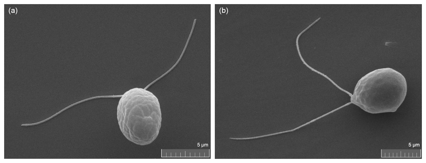
Figure 2Images taken of cryophilic microalgal species using a scanning electron microscope. WD = Working Distance, BI = Beam Intensity, MAG = Magnification. (a) Chlorominima collina cell. The microalgal cell has two flagella that it uses to swim. Culture ordered from the Culture Collection of Algae and Protozoa: CCAP 6/1. Strain isolated from Collins Glacier, King George Island, South Shetland Islands. WD: 9.97 mm, BI: 7.00, SEM MAG: 12.7 kx. Photo credit: Caitlin de Vries. (b) Limnomonas sp. cell. The microalgal cell has two flagella that it uses to swim. Culture ordered from the Culture Collection of Algae and Protozoa: CCAP 6/3 (Davey et al., 2019). Strain isolated from Rothera Point, Ryder Bay, Adelaide Island, Antarctic Peninsula. WD: 10.00 mm, BI: 7.00, SEM MAG: 10.5 kx. Photo credit: Caitlin de Vries.
Motile algal cells of the same species can be identified by the distinct gait that they exhibit due to factors such as the symmetry of their flagella and the fibres which attach the flagella to the basal body (Wan and Goldstein, 2016). For example, the model species Cd. reinhardtii has two physically symmetric flagella. The flagella's basal apparatus is structured so that it results in a non-planar, three-dimensional, flagellar beat (Wilson and Bees, 2025). Flagellar beating has been proven to be critical to phototactic response in Cd. reinhardtii (Wang et al., 2026) and is hypothesized to be relevant to other tactic responses, although this has not yet been proven. This, combined with the flagella having different phases, controls Cd. reinhardtii's helical swimming pattern (Cortese and Wan, 2021). Microalgae can also have morphologically asymmetric flagella. Golden snow algae from the genus Hydrurus such as H. nivalis and H. svalbardensis have physically asymmetric flagella, one long and one very short, only a fraction of the size of the other (Procházková et al., 2026), though little is known about Hydrurus's swimming pattern as many of the species have only recently been characterised.
This motility enables the microalgae to navigate complex environments such as snow, where their movement is heavily influenced by fluid dynamics, interactions with the medium's structure, and external stimuli. Understanding microalgal movement therefore requires consideration of the hydrodynamic regime within which microalgae operate.
In fluid mechanics, the Reynolds number (Re) is used as a dimensionless parameter to characterise the relative importance of inertial and viscous forces and to predict whether a flow will be laminar (viscous forces are dominant, Re ≪1) or turbulent (inertial forces are dominant, Re ≫1). For swimming microalgae in a fluid, Reynolds numbers are extremely low due to their small size, meaning that inertial forces are negligible compared to viscous forces. A Reynolds number is denoted by:
where ρ is fluid density, L is the characteristic length of the flow, which one can assume is the length of the algal cell body, μ the dynamic viscosity of the fluid and u0 is the swimming or flow speed.
For a contextual example using the model algal species Cd. reinhardtii, assuming a mean swimming speed of u0≈130 µm s−1 (Guasto et al., 2010), a cell length L≈10 µm (Marshall, 2024), water density ρ≈1000 kg m−3 and dynamic viscosity kg m−1 s−1, the Reynolds number is calculated as:
For an average human swimmer in the same environment (L≈1.7 m, ):
The Reynolds number corresponding to the human swimmer is roughly 109 times larger than that of the alga. In the case of the alga, viscous forces dominate and for the human swimmer, inertial forces dominate (Freund et al., 2012). The high ratio of viscous to inertial forces makes it difficult for microalgae to use the symmetrical swimming strategies employed by larger organisms like humans. For swimmers with low Reynolds numbers like microalgae, due to negligible inertial forces, reciprocal or symmetrical strokes are rendered ineffective for propulsion and produce no net motion. Instead, algae have evolved unique swimming techniques, such as the use of a helical flagellum and whip-like motions to navigate their environments effectively (Elgeti et al., 2015).
3.1 Snow
The structure of a snowpack, shaped by the arrangement and size of individual snow crystals, plays a key role in determining how motile algae navigate and colonise its interstitial spaces. Snow crystals primarily form when water vapor condenses onto tiny foreign particles, typically around one micron in size, known as cloud condensation nuclei, which include dust, sea salt, soot, pollen, and bacteria (Sturm, 2020). This process occurs in a supersaturated atmosphere. Although the unit cell structure of ice is tetrahedral due to the 104.5° angle of oxygen-hydrogen covalent bonding at the molecular level, snow crystal's hexagonal symmetry arises from the way the molecules stack together in a repeating pattern, creating a six-sided crystal lattice. Snow crystals will take on different morphologies e.g. dendrites, plates, needles or columns depending on the climatic and meteorological conditions under which they grow (Libbrecht, 2001).
Snow crystals fall to the ground and accumulate to form snowpacks. Within these snowpacks, the crystals undergo continuous metamorphic changes driven by variations in pressure and temperature gradients. As a result, the structure of the snowpack evolves over time and varies spatially throughout its depth. During this process, bonds develop between individual snow crystals through the exchange of water between vapour and solid phases. Low temperature gradients produce rounded snow crystals when the temperature gradient is °C per 10 cm, while high gradients °C per 10 cm create faceted ones (Libbrecht, 2019; Srivastava et al., 2010). Rounded crystals are commonly found in the upper portions of the snowpack. At the base, specifically under high temperature gradient conditions, repeated sublimation and recrystallisation will form large depth hoar crystals. In melting snow, where snow algae thrive, water films form around and between crystals, binding them into clusters.
Common snow algae species such as C. typhlos, Chl. collina, S. nivaloides and S. aurantia fall into a size range of approximately 5–40 µm in length (Gálvez et al., 2021; Procházková et al., 2019; Remias et al., 2005). Individual snow crystals typically range from < 0.5 to 3 mm, depending on crystal type (e.g. rounded crystals are smaller, while depth hoar crystals are larger) (Ishizaka, 1993; Mätzler, 2002), making snow crystals up to three orders of magnitude larger than the microalgae navigating around them, as seen in Fig. 3.
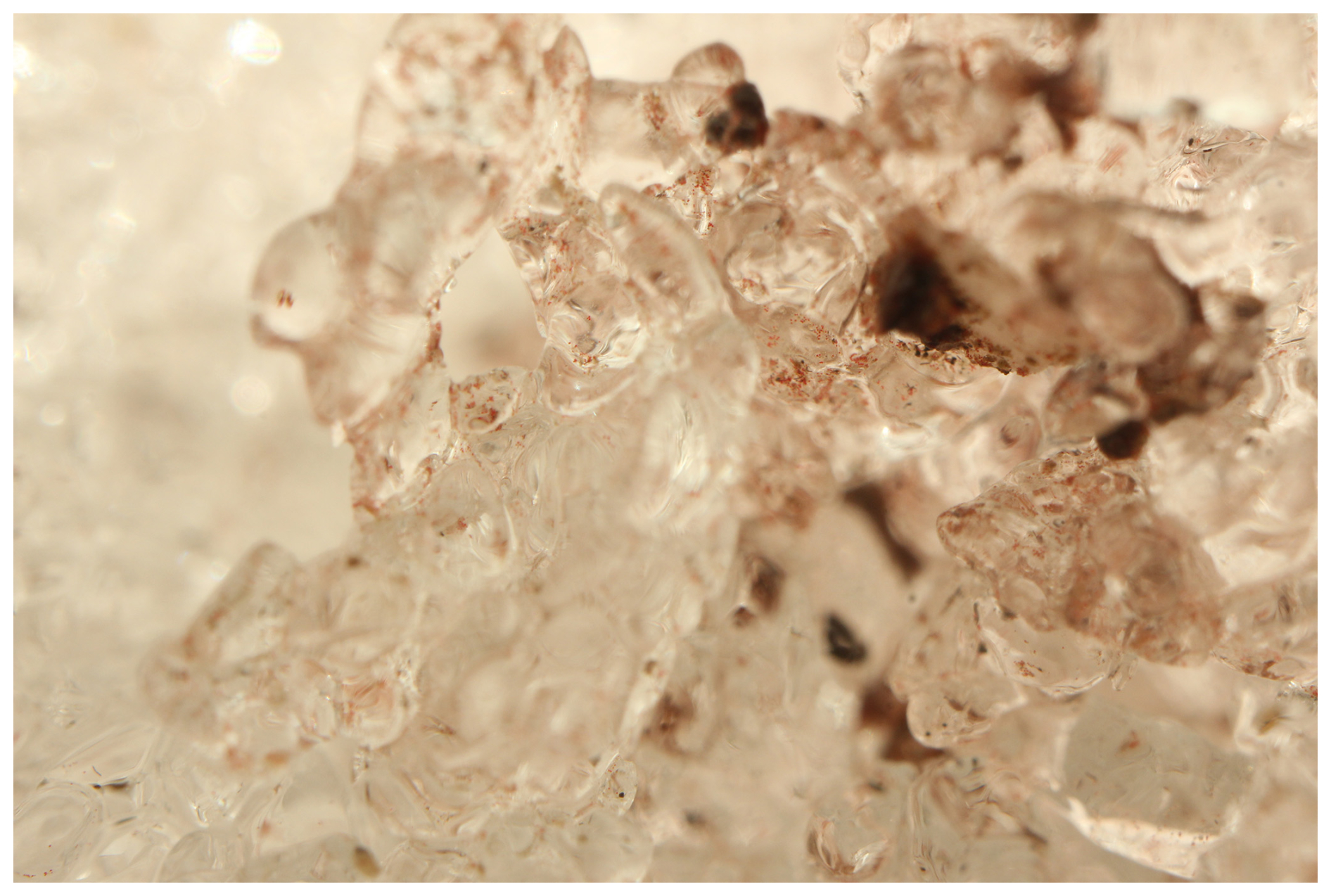
Figure 3In-situ photography of red snow algae cells within the snowpack, Robert Island, Antarctica (Thomson et al., 2025). Photo credit: Andrew Gray.
Fresh snow reflects more than 90 % of incoming visible radiation, making it the most reflective natural surface on Earth. Consequently, snow albedo represents one of the cryosphere's most significant influences on Earth's climate (Almela et al., 2025). The albedo of freshly fallen snow is ∼0.85, a unit-less measure of the fraction of incident solar radiation reflected by a surface (i.e. the ratio of upwelling to downwelling short-wave radiation) (De Vrese et al., 2021), described by the equation:
where α is the albedo, S↑ is the upwelling (reflected) solar radiation, and S↓ is the downwelling (incoming) solar radiation (Sandells and Flocco, 2022). From this, the amount of solar radiation absorbed by the surface, Snet, is:
Radiative forcing associated with snow arises from changes in the surface energy balance caused by reductions in snow albedo. This occurs through enhanced absorption of solar irradiance as the snow surface is darkened by light-absorbing particles, including snow algae, dust, and black carbon (Skiles et al., 2018).
3.1.1 Microalgal migration in a snowpack at the macroscopic scale
At the macroscopic scale, microalgal migration within snowpacks reflects the combined influence of environmental gradients, dispersal processes, and seasonal dynamics, shaping both the spatial distribution and timing of blooms. An original overview by Hoham and Duval (2001) described snow algae participating in a cyclical process where they overwinter as dormant cysts on a summer snowpack surface and/or the soil-snowpack interface. Current knowledge suggests that with the onset of spring snow melt, the cysts germinate into green, motile flagellated cells which respond to spring meltwater, light and the subsequent release of nutrients by migrating upward toward the newly accumulated snow surface formed during autumn and winter, as shown in Fig. 4. At the snow surface, or from another environmental cue, the microalgae transform into non-motile cells once again, appearing in colours including green, yellow, gold, orange and red. There is evidence for this lifecycle in the Chloromonas genus (e.g. Rea and Dial, 2024; Schuler and Mikucki, 2023), as well as a similar lifecycle in the Chlainomonas genus (though habitat specific in a snowy lake environment) (Matsumoto et al., 2024), but it has been only theorised for other algae genera and species.
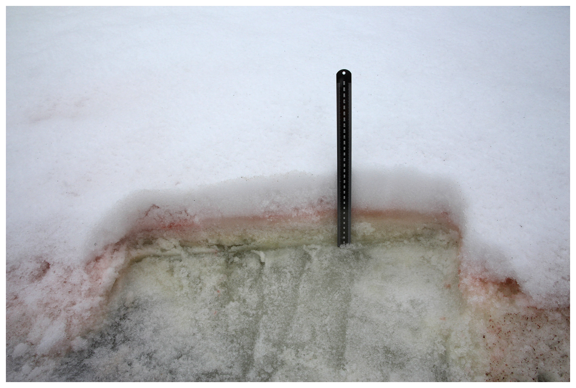
Figure 4Sub-surface green and red snow algae blooms, Ryder Bay, Antarctic Peninsula. Photo credit: Matthew Davey.
Snow algae have been shown to reduce snow albedo up to 44 % (Ganey et al., 2017; Lutz et al., 2016). Khan et al. (2021) quantified how differently coloured snow algal blooms influence snow albedo in the Antarctic Peninsula using field observations, spectral reflectance measurements, and pigment analysis. Compared to a control site without visible algae (0.85 ± 0.043), albedo was substantially reduced in algae-covered areas: 0.44 ± 0.12 for green-dominated sites, 0.65 ± 0.09 for red-dominated sites, and 0.58 ± 0.064 for mixed communities. This corresponds to an approximate 40 % reduction in snow albedo caused by green algal blooms and a 20 % reduction in snow albedo caused by red, with red communities also absorbing more light per unit pigment, particularly in the green wavelengths.
The temporal aspect of snow algal migration and lifecycle in Chlamydomonas and Chloromonas species has been described by Kvíderová (2010), having found snow algae at a study site in the Giant Mountains, Czech Republic to complete their entire lifecycle within a snowpack in a span of several weeks. Roussel et al. (2024) reported that the formation of red algal blooms (when snow algal cells reach their non-motile, red cyst phase on a snowpack surface) in the European Alps requires the presence of liquid water throughout the whole snow column for at least 46 d. One would hypothesise that this value would differ for differing species.
Liang et al. (2025) used high-resolution Sentinel-1 and Sentinel-2 satellite data to examine relationships between temperature, melt patterns, and snow algal biomass. Their multi-year analysis showed that algal biomass peaks about two months before maximum melt and temperature, likely because algae emerge early in the melt season when warming creates interstitial water for growth. The study also found that inconsistent intraseasonal temperatures hinder bloom development.
To investigate life cycle linked migration, during summer, Rea and Dial (2024) applied a bleach-containing mat to the surface of a subsection of an Alaskan ice field snowpack colonised by snow algae. Cell abundance beneath the mat, in the untreated areas surrounding the mat, and at a distant control site was measured the following summer after the treatment. The researchers defined two pathways for algae to recolonise the snow each year: active resurfacing and passive dispersal. Active resurfacing was hypothesised to occur when algal cysts germinate at the bottom of the snowpack entering the green motile phase in response to light and nutrient gradients, then transforming into non-motile, red cysts once again on top of the snowpack, dividing clonally (exhibiting mitosis and reproducing a genetically identical daughter cell). Passive dispersal was described as algal cells being introduced through passive transportation methods such as wind, water and birds. The authors concluded that at the peak of the growing season actively resurfacing cells were responsible for 65 % of microalgal surface abundance and that passive dispersion accounted for the remaining 35 %. The authors stated that no effort was made to classify the cells beyond being members of the Chlamydomonadaceae family.
These findings are consistent with Roussel et al. (2024), where Sentinel-2 satellite data from the European Alps displayed red algal blooms that persisted in environments where the ground was not permanently frozen. This pattern is notable in light of evidence that S. nivaloides cysts irreversibly lose photosynthetic capacity when exposed to −5 °C (Ezzedine et al., 2023; Roussel et al., 2024). Although the relationship remains speculative, inhibited photosynthetic ability would presumably impact upwards swimming behaviour motivated by phototaxis. If the active resurfacing pathway is curtailed, bloom dynamics should be similarly impacted.
3.1.2 Microalgal migration in a snowpack at the microscopic scale
On a microscopic scale, snow algae have been thought to inhabit the quasi-liquid layer (Grinde, 1983), a thin film of liquid water that surrounds snow crystals and persists at sub-zero temperatures, forming on the crystal surface within approximately 20 Kelvin of the melting point (Yasuda et al., 2024). This naturally raises the question of whether algae are able to actively swim within this layer. Early observations that appeared to support this idea were reported by Wergin et al. (1996), who examined metamorphosed snow crystals transported from remote regions in the United States. They identified structures thought to be red snow algae spores located just beneath the uppermost water film on the crystals, which could be revealed by etching away a few microns of ice from the surface.
However, the physical structure of liquid water within snowpacks may limit this possibility. In non-melting snow, the liquid water content is typically around 4 %–5 %. Due to surface tension, approximately 80 % of this water is held in menisci which form at contact points between snow crystal grains (Fig. 5) (Brzoska et al., 1998). The remaining 20 % exists as a quasi-liquid layer coating the snow crystals.
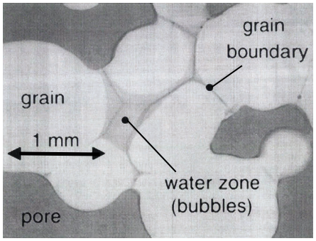
Figure 5A visualisation of the distribution of liquid water in snow with a high liquid water content, taken from Brzoska et al. (1998).
This quasi-liquid layer is extremely thin, ranging from only a few molecular layers (∼ 0.37 nm per layer; Sazaki et al., 2012) up to around 10 nm as temperature increases (Slater and Michaelides, 2019; Yasuda et al., 2024). In contrast, motile snow algae cells in their flagellated phase are several orders of magnitude larger, typically measuring 6–20 µm (Gálvez et al., 2021; Procházková et al., 2019; Remias et al., 2005), and up to 40 µm for species in some larger genera such as Chlainomonas (Matsumoto et al., 2024). The large size disparity suggests that active microalgal swimming behaviour within the quasi-liquid layer is unlikely. Instead, it may help explain why snow algae migration and bloom development tend to peak during snowmelt, when larger, interconnected water channels form that are sufficient to support cellular movement.
Ezzedine et al. (2023), using X-ray tomography at a µm resolution and focused-ion-beam scanning-electron-microscopy, observed field samples of S. nivaloides and found that dormant red cysts from this species were only present in the liquid water fraction of the snowpack, none appearing within the ice grain cores. These findings contrast with those of Ono and Takeuchi (2025), who reported that non-motile snow algal cysts were not transported through meltwater channels between snow crystals during daylight hours when snowmelt and channels of liquid water would have likely occurred. It is possible that Ono and Takeuchi (2025) were focused on migration at a macroscopic scale, with their most shallow samples being taken three centimetres from the snowpack surface, whereas Ezzedine et al. (2023) were doing up to a subcellular resolution of sample investigation. This could mean that there was potentially cell transport from percolation and cells present in liquid channels occurring in both studies, just not reported at a fine enough resolution for comparison purposes in the (Ono and Takeuchi, 2025) study. Additionally, the difference in findings could be related to potential melt and refreezing during sample transport during the Ezzedine et al. (2023) study. Or, lastly, the difference could also relate to the timescales of the studies or the environment that the microalgae were observed in, which could modify the microstructure of the snow, as we continue to discuss below.
Preferential flow paths within snowpacks form channels with flow velocities which have been observed to have speeds between 12 and 30 mm s−1 (Wakahama, 1968). The now polyphyletic snow alga Cd. nivalis, has a reported mean swimming speed of 0.061 mm s−1, negligible in comparison (Hill and Häder, 1997). However, snowpacks may not always support flow.
The characteristic viscous diffusion timescale τ, which determines how quickly momentum diffuses across a channel of width L, is given by:
where L is the width of a channel of flowing liquid within the snowpack (Fig. 6) and ν is the kinematic viscosity of the melting snow and ice. Here , where μ is the dynamic viscosity of the melting snow and ice and ρ is its density.
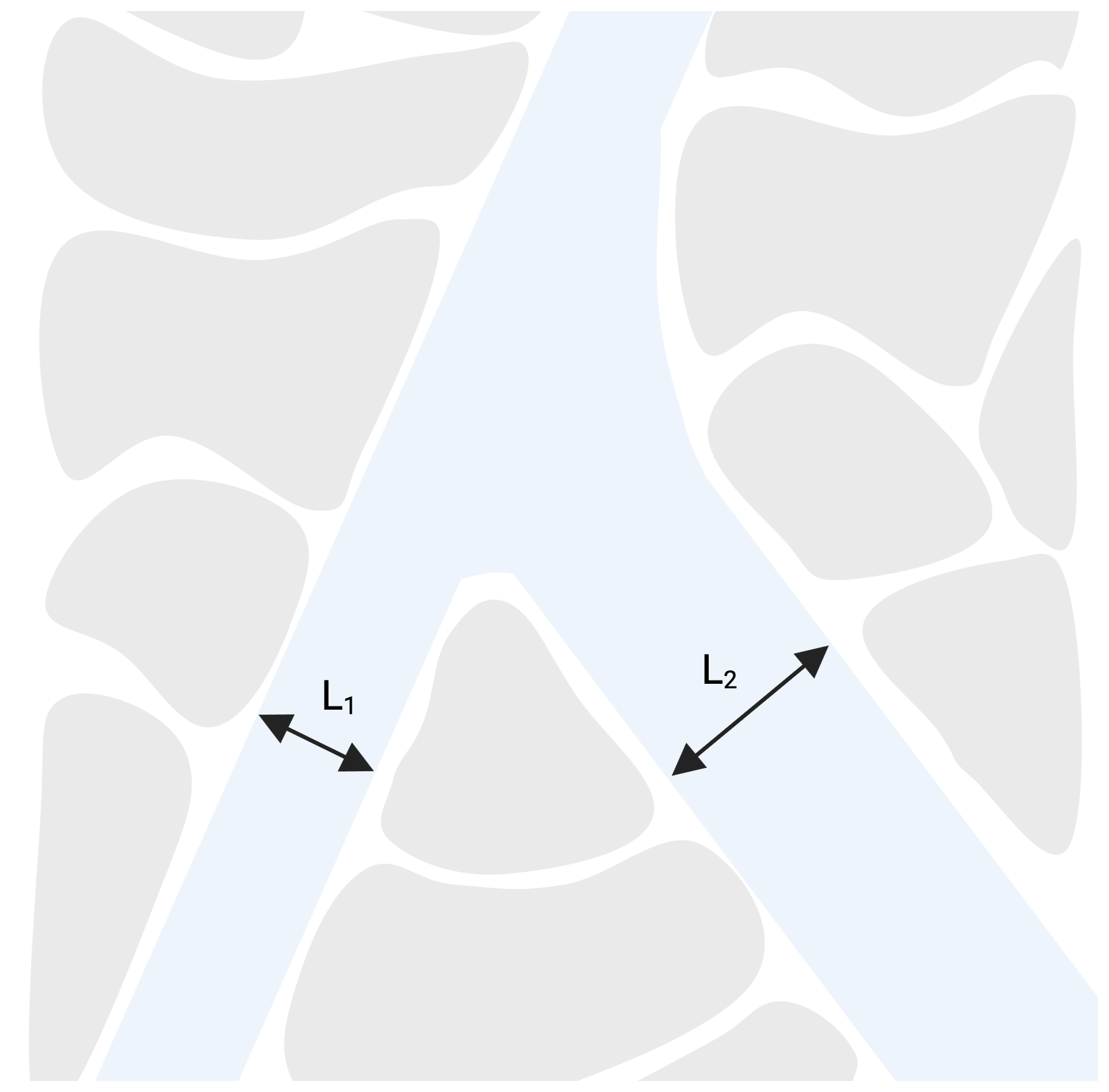
Figure 6A diagram of water channels within a snowpack, where L1 < L2. Water flowing in the narrower channel (L1) has a shorter viscous diffusion timescale than water flowing in the wider channel (L2). Created in BioRender (de Vries et al., 2026a).
This quantity measures how quickly momentum is transported through the action of viscosity from the boundaries towards the centre of a channel. This indicates that in narrow channels or high-viscosity regions, the timescale τ can be long, and snowpack flow may be effectively stagnant on the timescale of algal swimming. Wider channels are able to support sustained, fast-moving water, as momentum diffusion from the boundaries occurs over short timescales. Consequently, microalgae within these channels are likely to be passively transported by advection rather than actively swimming against the flow (however, swimming may still play a role due to cell rotation by shear and gravity, see Sect. 4.4 on gyrotaxis). When snow becomes fully saturated with liquid water, such as in capillary zones that form above impermeable layers like ice, water movement becomes negligible as τ is very small (τ∼1 s for a 100 µm-wide channel filled with water). These zones can reach thicknesses of up to approximately 20 cm (Coléou et al., 1999), providing an environment in which microalgae can move independently of bulk water flow (Bischoff, 2007). However, the limited thickness of these saturated regions suggests that additional transport must explain the presence of snow algae at greater heights within deeper snowpacks (Schuler and Mikucki, 2023).
Snow algae can also enhance their opportunity for migration through interfacial pre-melting. Interfacial pre-melting occurs when an active particle or organism is embedded within a host solid, such as ice, near its bulk melting temperature, where surface intermolecular forces induce the formation of a thin melted film at the interface of the particle and the host (Baran et al., 2025; Vachier and Wettlaufer, 2022). The thickness of the film around the active particle (e.g. snow algae) is dependent on temperature, impurities, material properties and geometry. This pre-melting also contributes to thermal regelation, which is the lowering of the melting point of a substance under pressure, and refreezing once this pressure is reduced. For example, as the ice or snow pre-melts against the algal cell, the imposition of a temperature gradient will cause the particle to move by a process of melting and refreezing. The alga will continue migrating towards comparatively warmer areas in the solid due to pressure differences caused by temperature gradient, the warmer areas experiencing lower pressure (Kreimer, 2009). Active particles such as biota have also developed unique survival strategies when trapped in ice, such as producing exopolymeric substances and antifreeze glycoproteins which increase interfacial melting, enhancing their motility and survival ability in harsh, icy conditions e.g. Meiners et al. (2003); Riedel et al. (2006). It has also been shown that motile microalgae in the genus Chlamydomonas can produce ice-binding proteins (Raymond and Morgan-Kiss, 2017), which bind to the surfaces of ice crystals and hinder their growth (Bar Dolev et al., 2016). Particle bio-locomotion can also be directed by nutrient availability in ice and snowpacks. Therefore, bio-enhanced thermal regelation and chemically directed bio-locomotion work together to govern algal motility in ice and snow (Vachier and Wettlaufer, 2022).
Tactic behaviour is a bias in swimming direction towards or away from a stimulus of biological, chemical or physical origins. For snow algae, these taxes in combination govern the cell's movement. We can model this using an agent based model, following what has been done for other algal species (Ishikawa et al., 2025). From the perspective of a cell, the following are equations of motion describing the orientation dynamics influenced by taxes.
The algal cell position evolves according to:
where is the local velocity of any flow in the liquid layers within the snowpack, x(t)∈ℝ3 denotes the algal cell's position, p(t)∈ℝ3 is its orientation unit vector, and vs is the constant swimming speed.
The algal cell's change in orientation with respect to time is given by:
Here, the first term denotes gravitactic reorientation, with timescale B, towards the vertical direction denoted by the unit vector k∈ℝ3. We see that when k=p, there is no contribution to the reorientation due to gravitaxis: cells swim upward (Pedley and Kessler, 1992). The second term denotes phototactic reorientation. We write this in the general form of a linear combination of reorientation toward direction of the light, with intensity I, given by the unit vector and reorientation in response to the intensity gradient ∇I (Williams and Bees, 2011a). Here β1 and β2 are constants and α(I) is a general function of light intensity I. If β1=0, phototactic reorientation will stop when p is aligned with the intensity gradient, while if β2=0, cells will no longer reorient when p is aligned with . The third term in Eq. (6) models reorientation by a flow with vorticity ω∈ℝ3. For simplicity we here assume cells are spherical and ignore their ellipsoidal shape; the effect of the flow for nonspherical cells can be modelled using a rate of strain tensor (Pedley and Kessler, 1992). The combination of the first and third term is known as gyrotaxis, which will be described in Sect.4.4. The fourth term models reorientation by chemotaxis, expressed by a general vector function χ(c,∇c), where c is a concentration of chemoattractant/chemorepellant and ∇c is its gradient. Similarly, reorientation by thermotaxis, is given by η(T,∇T), where T is the temperature of the local environment and ∇T its gradient. These taxes are expressed as general vector functions because they have not been mathematically modelled and remain, to the best of our knowledge, not well understood in microalgae, snow or otherwise. Understanding them may, in analogy with other microorganisms, involve spatial or temporal sensing, memory and adaptation (Othmer et al., 2013; Tan and Chiam, 2018). The final term models the reorientation of the cell due to noise, e.g. due to stochasticity in the flagellar beat (Wan and Goldstein, 2016). Lastly, ξ(t)∈ℝ3 is a 3D Gaussian white noise vector.
In addition to the assumptions mentioned, we should highlight that the agent based modelling framework above assumes that there is no coupling between taxes. An alternative approach to modelling populations of swimming microalgae is via continuum models, where differential equations for the probability density P(x,t) of finding microalgae at position x and time t are solved to evaluate the distribution of algae in space and time. We will not review these models here, but we refer the reader to the literature: Desai and Ardekani (2017); Pedley (2026); Pedley and Kessler (1992).
The following subsections examine what is known of each of the taxes listed in Eq. (6).
4.1 Phototaxis
Phototaxis is the movement of organisms in response to light stimuli. Positive phototaxis occurs when an organism migrates towards a light source, and negative when away (Bendix, 1960). It is a key behavioural trait in many snow algae (Détain et al., 2025). One of the most studied algae with regards to phototaxis is the freshwater microalga Cd. reinhardtii, with a recent review covering the details of this species' phototactic behaviour (Ishikawa et al., 2025). Like other microalgae, some snow algae exhibit positive phototaxis under low to moderate light intensities, enabling movement toward light sources to optimise photosynthesis (Détain et al., 2025). However, when light intensity exceeds a critical threshold, negative phototaxis is exhibited. For example, Ono and Takeuchi (2025) observed motile snow algae at the snowpack surface switching to negative phototaxis when solar radiation reached approximately 170 W m−2. This shift to negative phototaxis likely serves as a protective response, allowing cells to avoid light-induced stress such as photooxidation – a process in which intense light and oxygen lead to the degradation of chlorophyll and cellular organelles (Cheloni and Slaveykova, 2018; Elgeti et al., 2015; Foster and Smyth, 1980).
Ciliate microalgae sense light using eyespots and channelrhodopsins (Fig. 7) (Kreimer, 2009; Sineshchekov et al., 2009). Photoreceptor activation modulates intracellular calcium currents, inducing asymmetric flagellar beating and thereby steering cell movement toward or away from light sources (Pivato and Ballottari, 2021). Cd. reinhardtii swims in a helical path, so that its eyespot experiences an alternation of light and shade, which is used to control swimming direction towards the light, see Fig. 7. Interestingly, some snow algae without a visible eyespot have been shown to exhibit phototaxis as well (e.g. C. hindakii in Détain et al., 2025). Détain et al. (2025) examined phototaxis under varied temperature conditions in snow algal species L. spitsbergensis, Gloeocystsis sp., S. nivaloides, C. hindakii, Chrysophyceae sp., and Chloromonas sp. using a 540 nm green LED at 40 on one side of a Petri dish. Three of the species (S. nivaloides, C. hindakii and Chloromonas sp.) did not have a visible eyespot. Despite this, C. hindakii still exhibited phototactic behaviour whereas S. nivaloides and Chloromonas sp. did not. The remaining species had visible eyespots and performed phototaxis. This study brings into question how certain snow species navigate light intensity in snowpacks and how cell-light interactions function without eyespots. In this context, Novis et al. (2024) described a new species of snow algae in the Chloromonas genus, C. fuhrii, which among other unique defining characteristics does not have an eyespot (or a stigma as referred to in this study). The two nearest relatives of C. fuhrii reported, C. cf. platystigma and C. muramotoi both have eyespots, suggesting that its loss was recent. The authors speculated that the higher frequency of eyespot absence in species such as those from the Chloromonas genus which live in snow versus other habitats is potentially due to the dynamic of response to light in snow, suggesting that cells may need to avoid confusing signals from light reflecting from numerous directions off snow crystals. Eyespots are helpful for phototactic precision but have been proven to be unnecessary for phototactic behaviour itself as shown using Chlamydomonas mutants with no eyespot (Morel-Laurens and Feinleib, 1983).
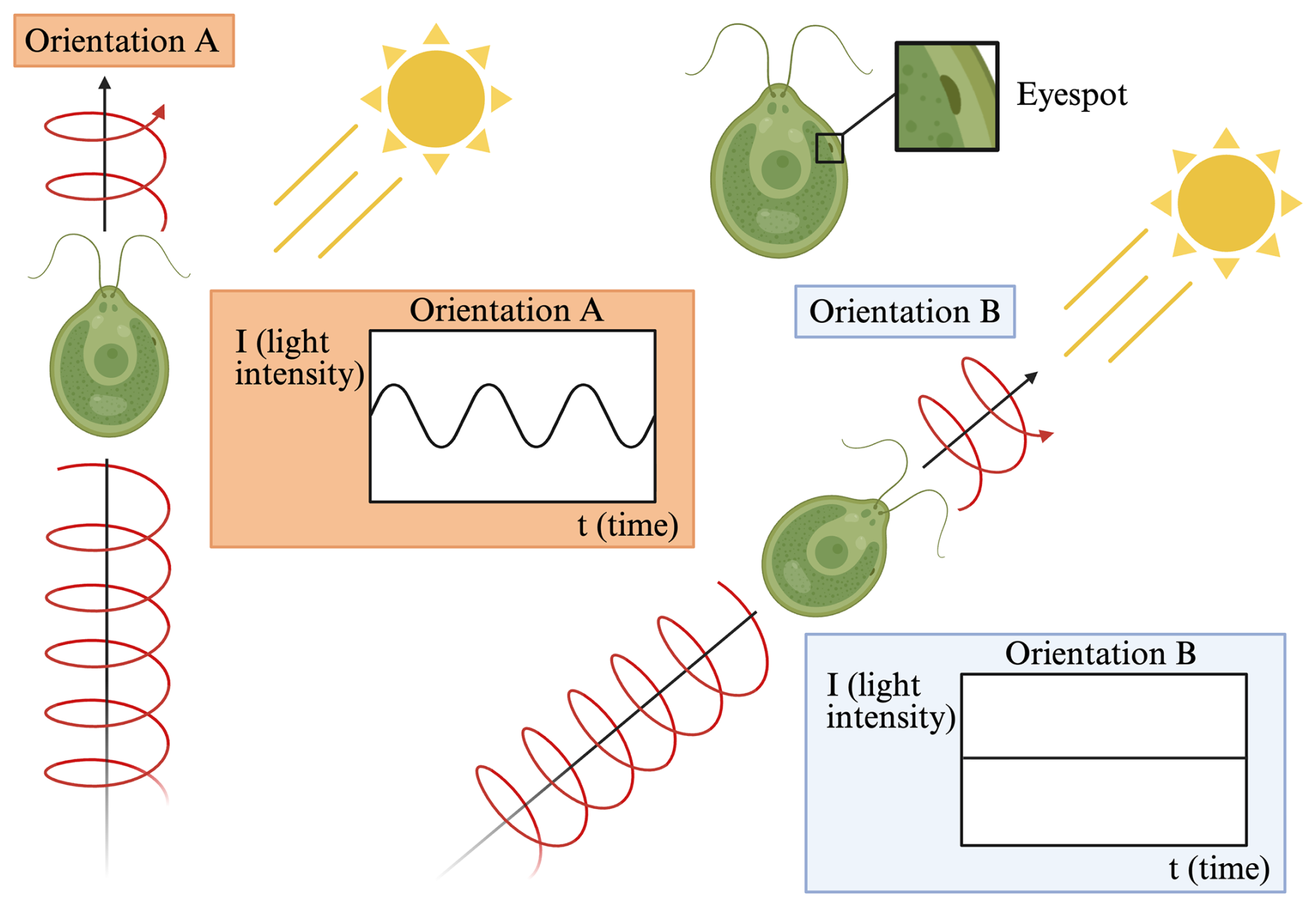
Figure 7Orientation A: A visualisation of a Chlamydomonas sp. cell swimming in a helical motion, oriented in a direction adjacent to a light source. As the cell swims helically (Cortese and Wan, 2021), the cell's singular eyespot rotates and perceives a sinusoidally varying light signal. Orientation B: A visualisation of a Chlamydomonas sp. cell swimming in a helical motion oriented towards a light source. The cell's singular eyespot experiences continuous, direct exposure to the light source. The time elapsed between orientation A and B is a few seconds (Choudhary et al., 2025; Goldstein, 2015). The time elapsed between orientation scenario A and B is negligible, otherwise one would see an increase in light intensity. Created in BioRender (de Vries et al., 2026b).
Ono and Takeuchi (2025) documented diurnal vertical migration of motile snow algae in an alpine forest snowpack in northern Japan. Motile algae ascended nearly to the snowpack surface towards nutrients and light in lower light hours and descended 10–20 cm into the snowpack during periods of peak solar radiation (a maximum of 755 W m−2) to avoid its intensity and remain at an optimal position for photosynthesis. Motile microalgal cell density at the surface layer was negatively correlated with solar radiation and air temperature (values ranging from R=−0.44 to −0.64, p<0.05); there was no correlation for non-motile microalgae. Despite solute and nutrient gradients throughout the snowpack, no day–night variation in solute distribution was detected, suggesting that light rather than nutrient availability drove migration. Additionally, algae have been shown to migrate away from predation by sensing infochemicals (kairomones) released from predators (Latta Iv et al., 2009). However, in the Ono and Takeuchi (2025) study, tardigrades and rotifers, which are potential snow algae predators, migrated before the snow algae when solar intensity increased, eliminating predator-induced chemotaxis as a potential driver of migration.
Häder and Häder (1989) described the effects of solar U.V.-B radiation on photo-orientation and motility in three flagellated species, including the now polyphyletic Cd. nivalis. Cd. nivalis, in its original growth medium, was exposed to solar radiation in two growth chambers: one with unfiltered sunlight and one with sunlight with U.V.-B cut-off filters and supplemented with ozone. Cd. nivalis exhibited high sensitivity to U.V.-B, although it did not demonstrate any clear phototactic orientation under unfiltered sunlight nor any initial positive light-induced swimming speed increase (photokinesis). After exposure, videotracking microscopy was used to quantify deviation from the stimulus direction. Under unfiltered light, after approximately 70 min of exposure, cell velocity quickly dropped and after approximately 90 min of exposure most cells were non motile. Motility stayed higher at all exposure times under the reduced U.V.-B radiation treatment. Motility increased with increasing filter wavelength, measured with 280, 295, 305 and 320 nm filters. The results suggest that UV-B acts mainly as a physiological stressor limiting motility, rather than a directional cue influencing phototaxis.
Algal phototaxis has also been demonstrated using Cd. reinhardtii under laboratory conditions in media containing a matrix of spherical obstacles (glass beads) (Prakash and Croze, 2021). The experiment, inspired by industrial purpose, attempted to maximise the quantity of microalgae at the medium's surface for ease of harvest. Cd. reinhardtii exhibited phototaxis under blue light () and the researchers were able to maximise algal density by incorporating 425–600 µm beads and a mesh with 350 µm pores within the liquid medium which significantly reduced bioconvective losses. This experiment provides valuable insight into how porous microstructures (as a proxy for snow crystals in snowpacks) can influence algal distribution in response to light gradients.
4.2 Chemotaxis
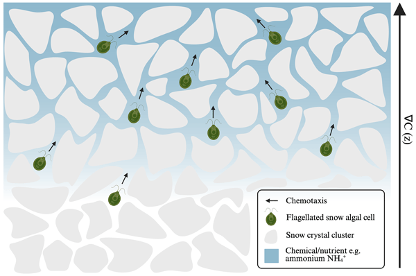
Chemotaxis is the directional movement of an organism along a chemical gradient, particularly in a positive direction towards nutrient sources (Adler et al., 1973) (Fig. 8) or in a negative direction away from toxic compounds (Young and Mitchell, 1973). Chemotaxis is also demonstrated in mating and predation scenarios (Govorunova and Sineshchekov, 2005; Latta Iv et al., 2009). Microalgae use receptors in their cell membrane to detect chemical cues, including ions and organic compounds, present in their surrounding environment (Amaral et al., 2023).
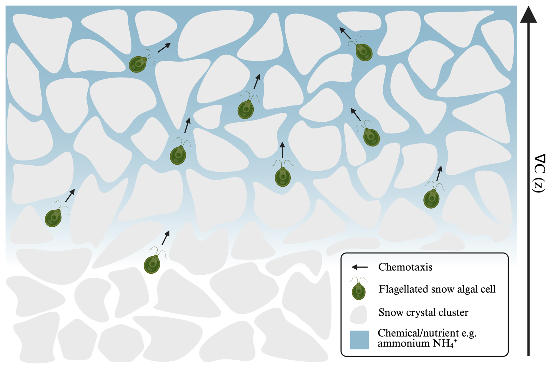
Figure 8A visualisation (not to scale) of snow algae navigating through a subsection of a snowpack with a chemical gradient present, denoted by ∇C, caused by a source at the top of the snowpack, exhibiting positive chemotaxis as they swim towards the nutrient source in the direction of the gradient's increase. Created in BioRender (de Vries et al., 2026c).
Snow algae inhabit snowpacks which are classified as oligotrophic environments where essential nutrients are often scarce and unevenly distributed (Maccario et al., 2015). Since nutrients such as nitrogen and phosphorus are required for algal growth and metabolism (Stibal et al., 2009), their spatial distribution may influence the movement of motile snow algal cells. Chemotaxis therefore represents a potential mechanism by which snow algae could respond to localised nutrient gradients.
Phosphorus is a limiting nutrient for the snow alga C. typhlos, whereas nitrogen deposition is hypothesized to have a limited effect on bloom occurrence and size (Almela et al., 2024). This conclusion is based on a 38 d incubation experiment at 4.5 °C using 24 nitrogen-to-phosphorus (N : P) treatments representative of nutrient availability in snow. Maximum biomass occurred at N : P molar ratios of 4–7, indicating that phosphorus availability is more important for optimising C. typhlos growth, although blooms developed across a wide range of nutrient conditions. Snow algal nutrient preference will vary across species, environment and lifecycle phase. Motile microalgae have been shown to exhibit chemotaxis toward metabolically relevant compounds, such as the movement of Cd. reinhardtii towards bicarbonate (Choi et al., 2016) and ammonium/methylammonium (Ermilova et al., 2007; Nelson et al., 2023) gradients. Such responses suggest that chemical gradients may guide algal movement toward favourable growth conditions. Given the apparent importance of phosphorus for snow algal growth in C. typhlos, phosphorus gradients may represent a potential driver of chemotactic behaviour, although this possibility has not yet been directly tested in snow algae.
It is also worth noting that snow algae have demonstrated plasticity in response to the nutrient conditions available to them (Broadwell et al., 2023). For example, Broadwell et al. (2023) demonstrated considerable stoichiometric plasticity across multiple snow algal strains, with cellular C:N ratios varying substantially under nitrate concentrations representative of natural snowpacks. Despite reduced growth under nutrient-limited conditions, the algae maintained growth across a broad range of nitrate availabilities, suggesting an ability to acclimate to nutrient-poor environments rather than relying solely on movement towards more favourable conditions. This has particular implications for the portion of cells belonging to motile snow algal species which are immobile either temporarily due to lifecycle stage, environmental stressors or other reasons.
As mentioned in the phototaxis section, a study by Ono and Takeuchi (2025) described the migration of microalgae within a snowpack on a mountain in Japan in response to solar radiation and nutrients. Motile snow algae were shown to migrate 10–20 cm downwards into the snowpack during times of intense solar radiation, returning nearer to the snowpack surface outside of these periods, presumably to benefit from available nutrients. The upper 23 cm of the snowpack were divided into one 5×3 cm layer (layer I) and four subsequently deeper 5×5 cm layers (II, III, IV and V). The upper layers (I–III) were characterised by higher concentrations of bioavailable nutrients such as ammonium (), phosphate (), and potassium (K+); the surface layer contained the highest proportions of (23.5 %), (20.8 %), and K+ (19.8 %). In contrast, deeper layers (IV–V) showed lower concentrations of nutrients which support algal growth, with Cl− and Na+ accounting for more than 60 % of total solutes below 15 cm depth. Meltwater did not redistribute the nutrient ratios with respect to snowpack layers and there was no significant difference between the solute distribution in the daytime (09:00–17:00 Japan Standard Time (JST), UTC+9, solar radiation > 150 W m−2) and nighttime (Ono and Takeuchi, 2025). The phenomenon observed in this study reinforces previously documented knowledge that the level of influence of phototaxis exceeds that of chemotaxis, but in the absence of solar radiation chemotaxis allows for vital nutrient uptake.
Cd. reinhardtii has been shown to exhibit chemotaxis toward ammonium in vitro (Nelson et al., 2023). In a Petri dish assay containing two agarose blocks, a 0 mM sink and a 21 mM NH4Cl source, cells migrated toward the ammonium enriched block within three hours, demonstrating chemotactic movement along the nutrient gradient. Using a wild-type strain (typical form of the species) the authors demonstrated enhanced chemotactic responses under light exposure; curiously, two phototaxis-incompetent mutant strains (eye3-2 and ptx1) still exhibited normal chemotaxis, showing that at least in this genus, chemotaxis and phototaxis pathways are independent. The lengthscale of the chemical gradient l in these and other lab-based studies was ∼ 1–10 mm, while in the field study of Ono and Takeuchi (2025), it was ∼ 10 cm. Chemotactic sensing on the scale of a snowpack depth may not be possible if l≫D, where D is the diameter of the algae, as the algae need to be able to compare significantly different concentrations as they swim. However, it seems possible that the microalgae will be able to chemotax up or down local gradients with smaller lengthscales, as depicted in Fig. 8.
Another instance of chemotactic behaviour in the Chlamydomonas genus is the detection and migration of opposite mating type gametes (mt+, mt−) in Cd. allensowrthii which is driven by gradients of sexual pheromones. Gametes are haploid cells specialised for sexual reproduction which fuse to form a zygote, and in this species both mating types are motile; however, only the mt− gametes (ancestral male) exhibit chemotactic behaviour towards the mt+ (ancestral female). The ability to detect the pheromone is growth stage specific, with vegetative cells being unstimulated and gametes only becoming chemotactic after gametogenesis (i.e. the process by which vegetative cells develop into reproductive gamete cells) (Govorunova and Sineshchekov, 2005). Sexual reproduction can facilitate genetic diversity and adaptability under certain environmental conditions (Agrawal, 2012). Chemotaxis may therefore play an important role in snow algae life histories under conditions including high solar radiation, temperature extremes, and low nutrient availability.
4.3 Gravitaxis
Gravitaxis (also referred to as geotaxis) is a direct response to gravity resulting in either the upward (negative gravitaxis) or downward (positive gravitaxis) orientation of a motile cell (Bean, 1977; Pedley and Kessler, 1992; Ishikawa et al., 2025). The mechanism driving gravitaxis has long been debated, with evidence supporting both passive physical processes (Kessler, 1985) and active physiological sensing (Häder et al., 2017). Biological responses which can govern gravitaxis include the activation of a gravity “sensor” like the use of mechanosensitive channels which intake chemicals at certain locations in the cell body (Häder and Lebert, 2001) or sedimenting statoliths, of which most protists do not utilise in their gravity orientations due to their small sizes (Häder and Hemmersbach, 2018). The earliest passive explanation, the “bottom-heavy” hypothesis, posits that an asymmetrical mass distribution causes the cell to orient itself upwards like a buoy, an idea first suggested by Wager (1911) and later modelled mathematically by Kessler (1985). An alternative passive model proposes that a cell's geometry (cell body plus flagella) leads to reorientation through a process of “differential sedimentation” where differences in gravitational settling rates between the cell body and flagella, generate a torque that reorients the cell (O'Malley and Bees, 2012; Roberts, 2006).
In the passive framework, a bottom-heavy cell is reoriented by a combination of a gravitational torque, Tg, due to gravity and a viscous torque, Tv, corresponding to resistance to rotation by the viscous fluid, see Fig. 9. At low Reynolds numbers inertia is negligible and the net torque on the body must be zero. As shown in Pedley and Kessler (1992), this implies that the cell orientation p reorients according to
where, as previously, B represents the characteristic reorientation time due to graviatxis and k is a unit vector pointing upwards. To get an idea of how this equation allows to predict the gravitactic reorientation, we shall derive a simpler two-dimensional version. In this case, and , where θ is the angle measured from the upwards unit vector k as shown in Fig. 9. Thus, substituting p and k into Eq. (7) and noting , we obtain
Then, differentiating p with respect to t:
Equating components of Eqs. (8) and (9), gives the equation for the change in orientation angle due to gravitaxis:
We can solve this equation for the orientation to the vertical of a gravitactic swimmer in 2D. The equilibrium orientation, θeq, occurs when , that is when , the vertical upwards direction. We note that this passive model is deterministic, i.e. it does not account for random reorientaion of gravitactic swimmers, which can be modelled in three-dimensions by a rotational noise term, as in Eq. (6).
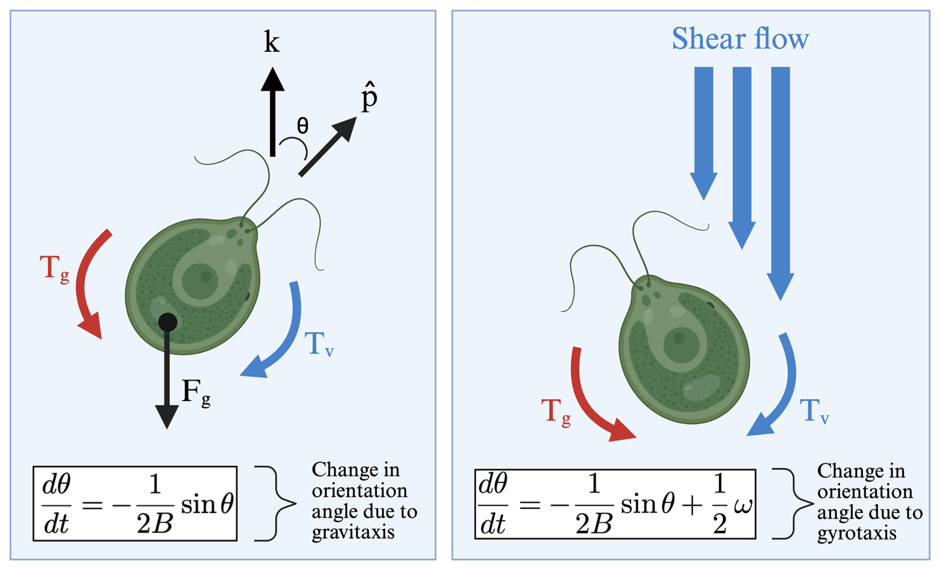
Figure 9Visualisation of change in orientation of a Chlamydomonas sp. cell due to gravitaxis and gyrotaxis. The diagram on the left represents pure gravitaxis: the cell body is reoriented by a torque Tg due to gravity and a resistive viscous torque Tv. B represents the characteristic reorientation time scale due to graviatxis, is a unit vector pointing in the direction of the cell's current orientation, k is the vertical unit vector defining the preferred upward direction for negative gravitaxis by convention, corresponding in two dimensions to θ=0. The diagram on the right represents gyrotaxis. Here the viscous torque also includes rotation by a shear flow with vorticity ω. As described in the main text, in two dimensions the preferred orientation with gyrotaxis is , that is the cell is oriented at an angle to the vertical. Created in BioRender (de Vries et al., 2026d).
It is still debated whether active or passive mechanisms are primarily responsible for gravitaxis in microalgae, appearing to be genera/species dependent as well as dependent on factors such as stress and age (Häder et al., 2017). Kam et al. (1999) examined gravitactic behaviour in Cd. reinhardtii, testing whether it relies on Ca2+ dependent pathways for gravitaxis. This was done using gadolinium and diltiazem which use different mechanisms to block a cell's Ca2+ channels, normally located at the cell's base and used as a mechanism for sensing orientation. It was found that the incorporation of neither chemical impacted Cd. reinhardtii's gravitational orientation, the population while exposed to said inhibitors still moved in a negatively gravitactic motion upwards, although gadolinium and diltiazem were found to reduce swimming speed. The phototaxis mutant ptx1 which, because of a defect in its flagellar apparatus, cannot reorient phototactically or chemotactically (Horst and Witman, 1993) exhibited normal gravitaxis as well. The authors concluded that Chlamydomonas's response to gravity is independent of calcium-mediated biochemical signal transduction and that calcium-mediated gravitaxis originated in an organism more “evolutionarily advanced”.
Evidence from mutant studies suggests that gravitaxis in Cd. reinhardtii is primarily an active, signal-transduction-driven process (Yoshimura et al., 2003). Yoshimura et al. investigated gravitaxis using a range of motility-, phototaxis-, and gravitaxis-related mutants. Mutants that swim only backwards (mbo1, mbo2) did not exhibit directed movement relative to gravity and instead sank at the same rate as non-motile cells, suggesting that passive factors such as cell density, shape, or random reorientation cannot fully explain gravitactic behaviour. Gravitaxis-deficient mutants (gtx1, gtx2) displayed normal motility but lacked gravitactic orientation, indicating that cells can swim normally yet fail to respond to gravitational cues. The authors further showed that impaired gravitaxis was associated with defects in membrane excitability, the ability of the cell membrane to undergo electrical changes through ion fluxes that regulate flagellar activity. For example, the phototaxis mutant ptx3, which has reduced membrane excitability, exhibited weakened gravitaxis, whereas ptx1, defective only in Ca2+-dependent flagellar dominance involved in phototaxis, retained normal gravitaxis. Together, these findings suggest that gravitaxis in Chlamydomonas relies on active physiological signalling rather than being solely a passive consequence of cell morphology or mass distribution.
Predictions derived from continuum and agent-based models which assume passive, torque driven reorientation of microalgae show good qualitative agreement with experimental observations (Barry et al., 2015; Sengupta et al., 2017). In cases where an alga lacks an active sensing mechanism, passive gravitactic reorientation provides a useful and informed initial approach for studying snow algae.
4.4 Gyrotaxis
When a microalgal cell is in a fluid flow it experiences a viscous torque caused by viscous drag, which is a drag force experienced by the organism due to the viscosity of the fluid surrounding it. Gyrotaxis refers to when the orientation of swimming microorganisms is governed by such viscous torques in combination with those due to gravity (Kessler, 1985; Pedley and Kessler, 1992).
Gyrotactic reorientation can be shown, similarly to the derivation of gravitaxis, to be given by (Pedley and Kessler, 1992)
where ω is the flow vorticity. Here for simplicity we neglect the effect of flow strain (Pedley and Kessler, 1992). To appreciate how Eq. (11) predicts gyrotactic cell orientation, we can consider two-dimensional dynamics of a swimmer in a 2D flow with vorticity , where ω is the magnitude of the z-component of the vorticity. In this case, recalling and noting that , following similar steps as for the derivation of Eq. (10), the reorientation rate of a gyrotactic swimmer is given by:
where θ is the orientation angle as measured from the vertical. We note that for gyrotaxis the equilibrium orientation is not necessarily upwards. By setting , Eq. (12) provides:
so that θeq=0 only in the absence of flow (ω=0). In general, cells swim at an angle to the vertical (θeq>0). We note that Eq. (13) is only satisfied for ωB≤1, outside these bounds, there is no equilibrium and cells will tumble rather that orderly orient (Pedley and Kessler, 1992). As for our discussion of gravitaxis, we have neglected reorientations due to rotational noise, which we included in Eq. (6). When these reorientations are included, gyrotactic cells still swim at an angle to the vertical in a shear flow, but this angle is not the one predicted by Eq. (13). The mean orientation can be predicted in 3D by advanced models beyond the scope of this review, see e.g. Fig. 1a, b of Bearon et al. (2012), which shows the components of the mean orientation of a gyrotactic swimmer.
Gyrotactic microorganisms are typically bottom-heavy, meaning their centre of mass lies below their geometric centre due to uneven internal mass distribution. This offset creates a gravitational torque when the cells swim horizontally, leading cells to swim upwards on average and accumulate at the surface of a suspension, leading to Rayleigh-Taylor instabilities when the motile organisms have a higher density than the surrounding media (Liu et al., 2020; Roberts, 2006; Vincent and Hill, 1996).
Bees and Hill (1997) investigated gyrotactic behaviour in bioconvective patterns formed by suspensions of the polyphyletic Cd. nivalis. To isolate gyrotaxis, phototactic responses were eliminated by illuminating the suspension from below with low-intensity red light, to which this species does not respond (Foster and Smyth, 1980). Under these conditions, pattern formation arose from the interaction between gyrotaxis and negative gravitaxis. Bioconvection occurs when biased swimming by motile microorganisms generates unstable cell concentration gradients, producing convective flows and spatial patterning within the suspension (Platt, 1961), as seen in Fig. 10.
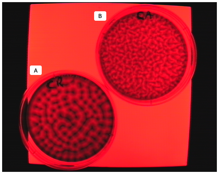
Figure 10Bioconvective patterns formed by microalgae. A: model mesophilic model species Cd. reinhardtii and B: snow algae C. typhlos in Petri dishes illuminated by deep red light (660 nm) to avoid a phototactic response (Foster and Smyth, 1980). Both the suspensions were well mixed prior to the spontaneous development of the patterns and had an average concentration ∼106 cells mL−1. Photo credit: Ottavio Croze.
The dynamics of gyrotactic orientation can be described using two key parameters: the cell reorientation time B which represents the characteristic time required for a cell to realign with gravity after being displaced, and the directional correlation time τ, which describes how long a cell maintains its swimming direction before random rotational diffusion causes it to lose memory of its initial orientation. Earlier theoretical work predicted that bioconvective instability occurs when these parameters are B≈ 1.25 s and τ≈ 5 s (Pedley and Kessler, 1990). The experimental Rayleigh numbers generated in the Bees and Hill (1997) study exceeded the critical threshold for instability predicted using these parameters. The results therefore provided experimental support for theoretical models of gyrotactic bioconvection, demonstrating that gyrotaxis alone can generate the regular, periodic bioconvective plumes observed (Bees and Hill, 1997).
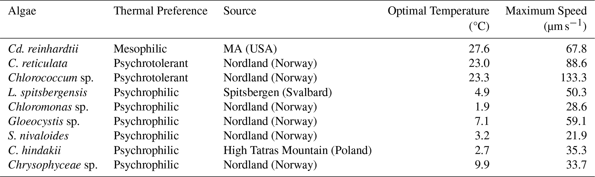
Table 1The specifications, optimal temperatures (for maximum swimming speed), the maximum speed and phototactic behaviour of select psychrophilic, psychrotolerant and mesophilic microalgae species. The microalgae were observed on a Peltier cooled microscope stage. Adapted from Détain et al. (2025).
In a later study, Williams and Bees (2011b) analysed the bioconvection patterns created by the gyrotactic snow algae Cd. augustae (previously listed as Cd. nivalis) in a well-mixed suspension under varied cell concentration and illumination scenarios. The phototactic, gyrotactic, and gravitactic responses were investigated during these instabilities using Fourier analysis to define pattern wavelength (the spacing between algal plumes in the bioconvective pattern) as a function of both cell concentration and light intensity. One of the experimental designs involved illuminating the algae suspension from above with white light, the most relevant arrangement for snow algae in a snowpack. The bioconvective pattern wavelength changed non-monotonically with light intensity. At low light intensities (645–1330 lux or ≈ 11–25 µmol m−2 s−1), cells exhibited strong phototaxis, swimming towards the light and reinforcing negative gravitaxis. The combined effect generated large overturning instabilities and broad, long wavelength patterns. Phototaxis weakened as intensity increased (1330–3000 lux or ≈22–56 µ). The orientational distribution of cells broadened due to weaker phototactic behaviour, making them more susceptible to viscous torques, so gyrotaxis became increasingly influential. Instabilities during this scenario were therefore less dominated by large overturning motion and instead began to show the influence of more localised, short-wavelength plumes. Near the critical intensity (3000 lux or ), cells near the top of the suspension exhibited negative phototaxis, swimming away from the intense light. Those lower in the suspension still swam upward as shading reduced the local intensity they experienced, creating a dense cell sublayer within the suspension where photo-gyrotactic instabilities produced small, fine-scale wavelength patterns. Outside of a laboratory environment, mathematical models assuming passive reorientation by gravity, have also made successful predictions for gyrotactic dispersion (Croze et al., 2017) and photogyrotactic bioconvection patterns (Williams and Bees, 2011a, b).
A final observation is that gyrotaxis may play a role in melting snow at the onset of the formation of draining flows through the snow. Cells advected by these downward flows may be gyrotactically focused toward the centre of the flow, where they drift down faster than the mean flow velocity (Croze et al., 2017). By this mechanism gyrotaxis may increase the transport of swimming algae to the bottom of the snowpack.
4.5 Thermotaxis and temperature sensitivity
Thermotaxis is the directional movement of an organism in response to a temperature gradient. Temperature-dependent motility (or thermokinesis) is when a motile cell has an ambient temperature range preference which governs its ability to demonstrate motility. Psychrophilic species of snow algae display maximum swimming speeds at colder temperatures than mesophilic species (Détain et al., 2025). To the best of our knowledge, no known studies on the thermotactic behaviour of snow algal species have been published, although limited literature exists on mesophilic species like Cd. reinhardtii (Sekiguchi et al., 2018).
Snow algal species are normally classified as psychrophilic, with an optimum growth range of 0–10 °C or psychrotolerant, capable of growing across a broader temperature range of up to ∼ 20 °C) (Broadwell et al., 2023; Gonzalez et al., 2026; Hoham and Remias, 2020). Détain et al. (2025) assessed the motility of six psychrophilic microalgal species/strains, two psychrotolerant and one mesophilic control under varied temperature conditions. The psychrophilic algae in this study included Limnomonas spitsbergensis, Chloromonas sp., Gleocystis sp., S. nivaloides, C. hindakii and, Chrysophyceae sp.. The psychrotolerant/mesophilic algae included Cd. reinhardtii, C. reticulata and Chlorococcum sp. (Table 1). Strong temperature-dependent motility responses were observed. All psychrophilic microalgae demonstrated an optimal temperature for maximum swimming speed of below 10 °C and all psychrotolerant and mesophilic algae demonstrated optimal temperatures for maximum swimming speed of above 20 °C.
Additionally, Sekiguchi et al. (2018) observed that the model alga Cd. reinhardtii responded to thermal gradients between 10 and 30 °, with the microalgae migrating towards lower temperatures regardless of the temperature that they were cultivated at. The thermotactic behaviour was due to membrane excitation and was governed by intracellular redox conditions (i.e. the cell's internal chemical state reflecting the balance of molecules that either gain or lose electrons, influencing stress and signaling pathways). The study showed that Cd. reinhardtii uses redox signals to adjust or even shut off its thermotactic behaviour, likely to prioritise other responses (such as phototaxis) when under conditions such as a lack of available nutrients or oxidative stress (Sekiguchi et al., 2018).
Recorded observations of snow algae navigating snowpacks in field and laboratory settings, macroscopically and microscopically, are limited in occurrence and understanding. Although the cyclical, seasonal process of snow algae overwintering as cysts and becoming flagellated to access light and nutrients is agreed upon, the interaction with quasi-liquid layers on snow crystals, navigation through meltwater channels, responses to snowpack evolution and behaviour under flow conditions have not been quantitatively documented from a biophysical perspective. There is a large knowledge gap surrounding the tactic behaviour of snow algae species, particularly concerning gyrotaxis and thermotaxis. Literature on phototaxis and chemotaxis largely pertains to genera that include some snow algae species (e.g. Chloromonas), rather than psychrophilic species specifically. Further developing equations describing the biomechanics of snow algae motility, informed by experimental results at single cell and population levels of resolution would enable deeper understanding and improved prediction of snow algal blooms and their drivers. This new knowledge could further the understanding of microswimmer behaviour in active matter physics, aid in the protection of vulnerable habitats and species, improve the accuracy of hydrological and cryospheric models, and contribute to advances in biotechnology and medicine.
No code was generated or used within this manuscript.
No data were generated or used within this manuscript.
CSdV: Conceptualisation, Visualisation, Writing – original draft, Writing – review & editing, MJS: Supervision, Writing – review & editing, MPD: Writing – review & editing, GSC: Writing – review & editing, OAC: Conceptualisation, Visualisation, Supervision, Writing – review and editing.
The contact author has declared that none of the authors has any competing interests.
Publisher's note: Copernicus Publications remains neutral with regard to jurisdictional claims made in the text, published maps, institutional affiliations, or any other geographical representation in this paper. The authors bear the ultimate responsibility for providing appropriate place names. Views expressed in the text are those of the authors and do not necessarily reflect the views of the publisher.
The high resolution images of snow algal cells included in this review were taken using the scanning electron microscopy facilities at Newcastle University. Illustration diagrams in this manuscript (Figs. 6, 7, 8 and 9) were created with BioRender.com (de Vries et al., 2026a).
The first author (CSdV) acknowledges funding from the Natural Environment Research Council (NERC) through a UK Research and Innovation (UKRI) Doctoral Training Partnership (NE/S007512/1). The third author (MPD) acknowledges support from a UKRI NERC grant (NE/V000764/1) and from UKRI CCAP facilities under grant (NE/Y006321/1).
This paper was edited by Susanne Liebner and reviewed by two anonymous referees.
Adler, J., Hazelbauer, G. L., and Dahl, M. M.: Chemotaxis toward sugars in Escherichia coli, J. Bacteriol., 115, 824–847, https://doi.org/10.1128/jb.115.3.824-847.1973, 1973. a
Agrawal, S. C.: Factors controlling induction of reproduction in algae – review: the text, Folia Microbiol., 57, 387–407, https://doi.org/10.1007/s12223-012-0147-0, 2012. a
Almela, P., Elser, J. J., Giersch, J. J., Hotaling, S., Rebbeck, V., and Hamilton, T. L.: Laboratory experiments suggest a limited impact of increased nitrogen deposition on snow algae blooms, Env. Microbiol. Rep., 16, e70 052, https://doi.org/10.1111/1758-2229.70052, 2024. a
Almela, P., Elser, J. J., Giersch, J. J., Hotaling, S., and Hamilton, T. L.: Influence of snow cover on albedo reduction by snow algae, mBio, 16, e03630-24, https://doi.org/10.1128/mbio.03630-24, 2025. a
Amaral, R., Duci, D., Cotta, F. C., Bacellar, F. L., Oliveira, S., Verret, F., Asadi, K., Vandamme, L. K., Reis, N. M., Bryant, L. D., Tosh, D., Mouget, J.-L., Perkins, R., and Rocha, P. R.: Ion-driven communication and acclimation strategies in microalgae, Chem. Eng. J., 473, 144985, https://doi.org/10.1016/j.cej.2023.144985, 2023. a
Bar Dolev, M., Braslavsky, I., and Davies, P. L.: Ice-Binding Proteins and Their Function, Annu. Rev. Biochem., 85, 515–542, https://doi.org/10.1146/annurev-biochem-060815-014546, 2016. a
Baran, Ł., Llombart, P., and MacDowell, L. G.: Understanding Interfacial Ice Premelting: Structure, Adhesion, and Nucleation, J, Phys. Chem. C, 129, 4614–4631, https://doi.org/10.1021/acs.jpcc.4c07328, 2025. a
Barry, M. T., Rusconi, R., Guasto, J. S., and Stocker, R.: Shear-induced orientational dynamics and spatial heterogeneity in suspensions of motile phytoplankton, J. Roy. Soc. Interface, 12, 20150791, https://doi.org/10.1098/rsif.2015.0791, 2015. a
Barsanti, L., Birindelli, L., Di Garbo, A., and Gualtieri, P.: Bioconvection in microalgae: review of mathematical models, Appl. Sci., 15, 2708, https://doi.org/10.3390/app15052708, 2025. a
Bean, B.: Geotactic behavior of Chlamydomonas, J. Protozool., 24, 394–401, https://doi.org/10.1111/j.1550-7408.1977.tb04759.x, 1977. a
Bearon, R. N., Bees, M. A., and Croze, O. A.: Biased swimming cells do not disperse in pipes as tracers: A population model based on microscale behaviour, Phys. Fluids, 24, 121902, https://doi.org/10.1063/1.4772189, 2012. a
Bechinger, C., Di Leonardo, R., Löwen, H., Reichhardt, C., Volpe, G., and Volpe, G.: Active particles in complex and crowded environments, Rev. Modern Phys., 88, 045006, https://doi.org/10.1103/RevModPhys.88.045006, 2016. a
Bees, M. A. and Hill, N. A.: Wavelengths of bioconvection patterns, J. Exp. Biol., 200, 1515–1526, https://doi.org/10.1242/jeb.200.10.1515, 1997. a, b, c
Bendix, S. W.: Phototaxis, Bot. Rev., 26, 145–208, https://doi.org/10.1007/BF02860529, 1960. a, b
Bischoff, Y.: Diversité et mobilitédes algues de neige dans les Alpes suisses, PhD thesis, Université de Genée, https://doi.org/10.13097/ARCHIVE-OUVERTE/UNIGE:516, 2007. a
Broadwell, E. L. M., Pickford, R. E., Perkins, R. G., Sgouridis, F., and Williamson, C. J.: Adaptation versus plastic responses to temperature, light, and nitrate availability in cultured snow algal strains, FEMS Microbiol. Ecol., 99, fiad088, https://doi.org/10.1093/femsec/fiad088, 2023. a, b, c
Brown, S. P., Olson, B. J., and Jumpponen, A.: Fungi and algae co-occur in snow: an issue of shared habitat or algal facilitation of heterotrophs?, Arct. Antarct. Alp. Res., 47, 729–749, https://doi.org/10.1657/AAAR0014-071, 2015. a
Brzoska, J.-B., Coléou, C., and Lesaffre, B.: Thin-sectioning of wet snow after flash-freezing, J. Glaciol., 44, 54–62, https://doi.org/10.3189/S0022143000002343, 1998. a, b
Cheloni, G. and Slaveykova, V.: Photo-Oxidative Stress in Green Algae and Cyanobacteria, Reactive Oxygen Species, https://archive-ouverte.unige.ch/unige:102840 (last access: 24 September 2026), 2018. a
Chen, D., Heymann, M., Fraden, S., Nicastro, D., and Dogic, Z.: ATP consumption of eukaryotic flagella measured at a single-cell level, Biophys. J., 109, 2562–2573, https://doi.org/10.1016/j.bpj.2015.11.003, 2015. a
Choi, H. I., Kim, J. Y. H., Kwak, H. S., Sung, Y. J., and Sim, S. J.: Quantitative analysis of the chemotaxis of a green alga, Chlamydomonas reinhardtii, to bicarbonate using diffusion-based microfluidic device, Biomicrofluidics, 10, 014121, https://doi.org/10.1063/1.4942756, 2016. a
Choudhary, S. K., Ma, Y., Baskaran, A., and Sharma, P.: Transient state dynamics of Chlamydomonas reinhardtii cells during phototaxis, Phys. Rev. Res., 7, 033282, https://doi.org/10.1103/l53n-2kg1, 2025. a
Coléou, C., Xu, K., Lesaffre, B., and Brzoska, J.-B.: Capillary rise in snow, Hydrol. Process., 13, 1721–1732, https://doi.org/10.1002/(SICI)1099-1085(199909)13:12/13<1721::AID-HYP852>3.0.CO;2-D, 1999. a
Cortese, D. and Wan, K. Y.: Control of Helical Navigation by Three-Dimensional Flagellar Beating, Phys. Rev. Lett., 126, 088003, https://doi.org/10.1103/PhysRevLett.126.088003, 2021. a, b
Croze, O. A., Bearon, R. N., and Bees, M. A.: Gyrotactic swimmer dispersion in pipe flow: testing the theory, J. Fluid Mech., 816, 481–506, https://doi.org/10.1017/jfm.2017.90, 2017. a, b
Davey, M. P., Norman, L., Sterk, P., Huete-Ortega, M., Bunbury, F., Loh, B. K. W., Stockton, S., Peck, L. S., Convey, P., Newsham, K. K., and Smith, A. G.: Snow algae communities in Antarctica: metabolic and taxonomic composition, New Phytol., 222, 1242–1255, https://doi.org/10.1111/nph.15701, 2019. a, b, c
Détain, A., Suzuki, H., Wijffels, R. H., Leborgne-Castel, N., and Hulatt, C. J.: Snow algae exhibit diverse motile behaviors and thermal responses, mBio, e02954-24, https://doi.org/10.1128/mbio.02954-24, 2025. a, b, c, d, e, f, g
De Vrese, P., Stacke, T., Caves Rugenstein, J., Goodman, J., and Brovkin, V.: Snowfall-albedo feedbacks could have led to deglaciation of snowball Earth starting from mid-latitudes, Commun. Earth Environ., 2, 91, https://doi.org/10.1038/s43247-021-00160-4, 2021. a
de Vries, C. S.: A diagram of water channels within a snowpack, where L1 < L2. Water flowing in the narrower channel (L1) has a shorter viscous diffusion timescale than water flowing in the wider channel (L2), https://BioRender.com/1oc2605 (last access: 5 August 2026), 2026a. a, b
de Vries, C. S.: Orientation A: A visualisation of a Chlamydomonas sp. cell swimming in a helical motion, oriented in a direction adjacent to a light source. As the cell swims helically (Cortese and Wan, 2021), the cell’s singular eyespot rotates and perceives a sinusoidally varying light signal. Orientation B: A visualisation of a Chlamydomonas sp. cell swimming in a helical motion oriented towards a light source. The cell’s singular eyespot experiences continuous, direct exposure to the light source. The time elapsed between orientation A and B is a few seconds (Choudhary et al., 2025; Goldstein, 2015). The time elapsed between orientation scenario A and B is negligible, otherwise one would see an increase in light intensity, https://BioRender.com/r06ua93 (last access: 5 August 2026), 2026b. a
de Vries, C. S.: A visualisation (not to scale) of snow algae navigating through a subsection of a snowpack with a chemical gradient present, denoted by ∇C, caused by a source at the top of the snowpack, exhibiting positive chemotaxis as they swim towards the nutrient source in the direction of the gradient’s increase, https://BioRender.com/inc0hfl (last access: 5 August 2026), 2026c. a
de Vries, C. S.: A two-dimensional (for simplicity) visualisation of change in orientation of a Chlamydomonas sp. cell due to gravitaxis and flow of surrounding medium. The diagram on the left represents pure gravitaxis: the cell body is reoriented by a torque Tg due to gravity and a resistive viscous torque Tv. B represents the characteristic reorientation time scale due to graviatxis, ∧p is a unit vector pointing in the direction of the cell’s current orientation, k is the vertical unit vector defining the preferred upward direction for negative gravitaxis by convention, corresponding in 2D to θ=0. The diagram on the right represents gyrotaxis. Here the viscous torque also includes rotation by a shear flow with vorticity ω. The prefereed orientation is now at an angle, θ=θc, to the vertical, https://BioRender.com/duylke2 (last access: 5 August 2026), 2026d. a
Desai, N. and Ardekani, A. M.: Modeling of active swimmer suspensions and their interactions with the environment, Soft Matter, 13, 6033–6050, https://doi.org/10.1039/C7SM00766C, 2017. a
Duval, B., Shetty, K., and Thomas, W. H.: Phenolic compounds and antioxidant properties in the snow alga Chlamydomonas nivalis after exposure to UV light, J. Appl. Phycol., 11, 559–566, https://doi.org/10.1023/A:1008178208949, 1999. a, b
Ebbens, S. J. and Gregory, D. A.: Catalytic janus colloids: controlling trajectories of chemical microswimmers, Accounts Chem. Res., 51, 1931–1939, https://doi.org/10.1021/acs.accounts.8b00243, 2018. a
Elgeti, J., Winkler, R. G., and Gompper, G.: Physics of microswimmers – single particle motion and collective behavior: A review, Rep. Prog. Phys., 78, 056601, https://doi.org/10.1088/0034-4885/78/5/056601, 2015. a, b, c
Engstrom, C. B., Raymond, B. B., Albeitshawish, J., Bogdanovic, A., and Quarmby, L. M.: Rosetta gen. nov. (Chlorophyta): Resolving the identity of red snow algal rosettes, J. Phycol., 60, 275–298, https://doi.org/10.1111/jpy.13438, 2024. a, b
Ermilova, E. V., Nikitin, M. M., and Fernández, E.: Chemotaxis to ammonium/methylammonium in Chlamydomonas reinhardtii: the role of transport systems for ammonium/methylammonium, Planta, 226, 1323–1332, https://doi.org/10.1007/s00425-007-0568-1, 2007. a
Ezzedine, J. A., Uwizeye, C., Si Larbi, G., Villain, G., Louwagie, M., Schilling, M., Hagenmuller, P., Gallet, B., Stewart, A., Petroutsos, D., Devime, F., Salze, P., Liger, L., Jouhet, J., Dumont, M., Ravanel, S., Amato, A., Valay, J.-G., Jouneau, P.-H., Falconet, D., and Maréchal, E.: Adaptive traits of cysts of the snow alga Sanguina nivaloides unveiled by 3D subcellular imaging, Nat. Commun., 14, 7500, https://doi.org/10.1038/s41467-023-43030-7, 2023. a, b, c, d, e
Foster, K. W. and Smyth, R. D.: Light antennas in phototactic algae, Microbiol. Rev., 44, 572–630, https://doi.org/10.1128/mr.44.4.572-630.1980, 1980. a, b, c
Freund, J. B., Goetz, J. G., Hill, K. L., and Vermot, J.: Fluid flows and forces in development: functions, features and biophysical principles, Development, 139, 1229–1245, https://doi.org/10.1242/dev.073593, 2012. a
Gálvez, F. E., Saldarriaga-Córdoba, M., Huovinen, P., Silva, A. X., and Gómez, I.: Revealing the characteristics of the Antarctic snow alga Chlorominima collina gen. et sp. nov. through taxonomy, physiology, and transcriptomics, Front. Plant Sci., 12, 662298, https://doi.org/10.3389/fpls.2021.662298, 2021. a, b
Ganey, G. Q., Loso, M. G., Burgess, A. B., and Dial, R. J.: The role of microbes in snowmelt and radiative forcing on an Alaskan icefield, Nat. Geosci., 10, 754–759, https://doi.org/10.1038/ngeo3027, 2017. a, b
Ganguly, A., Alessio, B. M., and Gupta, A.: Diffusiophoresis: a novel transport mechanism – fundamentals, applications, and future opportunities, Frontiers in Sensors, 4, 1322906, https://doi.org/10.3389/fsens.2023.1322906, 2023. a
Goldstein, R. E.: Green algae as model organisms for biological fluid dynamics, Annu. Rev. Fluid Mech., 47, 343–375, https://doi.org/10.1146/annurev-fluid-010313-141426, 2015. a
Gonzalez, C. R., Grigg, V., Thomas, N., Thomson, A. I., MacPherson, E., Dillon, L., Gober, E., Smith, A. G., Cockell, C., Convey, P., and Davey, M. P.: Antarctic snow algal responses to temperature – potential implications of climate change, Sustainable Microbiology, 3, qvag023, https://doi.org/10.1093/sumbio/qvag023, 2026. a, b
Wakahama, G.: The metamorphism of wet snow, in: General Assembly of Bern 1967 – Snow and Ice, vol. 79 of International Association of Scientific Hydrology Publication, 370–379, https://iahs.info/uploads/dms/079035.pdf (last access: 24 September 2026), 1968. a
Gorton, H. L., Williams, W. E., and Vogelmann, T. C.: The Light Environment and Cellular Optics of the Snow Alga Chlamydomonas nivalis (Bauer) Wille, Photochem. Photobiol., 73, 611–620, https://doi.org/10.1562/0031-8655(2001)0730611TLEACO2.0.CO2, 2007. a
Govorunova, E. G. and Sineshchekov, O. A.: Chemotaxis in the green flagellate alga Chlamydomonas, Biochemistry (Moscow), 70, 717–725, https://doi.org/10.1007/s10541-005-0176-2, 2005. a, b
Gray, A., Krolikowski, M., Fretwell, P., Convey, P., Peck, L. S., Mendelova, M., Smith, A. G., and Davey, M. P.: Remote sensing reveals Antarctic green snow algae as important terrestrial carbon sink, Nat. Commun., 11, 2527, https://doi.org/10.1038/s41467-020-16018-w, 2020. a
Grinde, B.: Vertical distribution of the snow alga Chlamydomonas nivalis (Chlorophyta, Volvocales), Polar Biol., 2, 159–162, https://doi.org/10.1007/BF00448965, 1983. a
Guasto, J. S., Johnson, K. A., and Gollub, J. P.: Measuring Oscillatory Velocity Fields Due to Swimming Algae, arXiv [preprint], https://doi.org/10.48550/arxiv.1010.1787, 2010. a
Häder, D.-P. and Hemmersbach, R.: Gravitaxis in flagellates and ciliates, in: Gravitational Biology I, Springer International Publishing, Cham, 27–45, ISBN 978-3-319-93893-6 978-3-319-93894-3, https://doi.org/10.1007/978-3-319-93894-3_3, 2018. a
Häder, D.-P. and Häder, M. A.: Effects of solar u.v.-B irradiation on photomovement and motility in photosynthetic and colorless flagellates, Environ. Exp. Bot., 29, 273–282, https://doi.org/10.1016/0098-8472(89)90059-2, 1989. a
Häder, D.-P. and Lebert, M.: Graviperception and gravitaxis in algae, Adv. Space Res., 27, 861–870, https://doi.org/10.1016/S0273-1177(01)00149-1, 2001. a, b
Häder, D.-P., Braun, M., Grimm, D., and Hemmersbach, R.: Gravireceptors in eukaryotes – a comparison of case studies on the cellular level, npj Microgravity, 3, 13, https://doi.org/10.1038/s41526-017-0018-8, 2017. a, b
Hill, N. and Häder, D.-P.: A biased random walk model for the trajectories of swimming micro-organisms, J. Theor. Biol., 186, 503–526, https://doi.org/10.1006/jtbi.1997.0421, 1997. a
Hoham, R. W.: Optimum Temperatures and Temperature Ranges for Growth of Snow Algae, Arct. Alp. Res., 7, 13–24, https://doi.org/10.1080/00040851.1975.12003805, 1975. a
Hoham, R. W. and Duval, B.: Snow ecology: an interdisciplinary examination of snow-covered ecosystems, in: Snow Ecology: An Interdisciplinary Examination of Snow-Covered Ecosystems, Cambridge University Press, Cambridge, 168–228, ISBN13: 9780521188890, ISBN10: 052118889X, 2001. a
Hoham, R. W. and Ling, H. U.: Snow Algae: The Effects of Chemical and Physical Factors on Their Life Cycles and Populations, in: Journey to Diverse Microbial Worlds, edited by Seckbach, J., Springer Netherlands, Dordrecht, 131–145, ISBN 978-94-010-5850-6 978-94-011-4269-4, https://doi.org/10.1007/978-94-011-4269-4_10, 2000. a
Hoham, R. W. and Mullet, J. E.: The life history and ecology of the snow alga Chloromonas cryophila sp. nov. (Chlorophyta, Volvocales), Phycologia, 16, 53–68, https://doi.org/10.2216/i0031-8884-16-1-53.1, 1977. a
Hoham, R. W. and Remias, D.: Snow and glacial algae: A review, J. Phycol., 56, 264–282, https://doi.org/10.1111/jpy.12952, 2020. a, b, c
Horst, C. J. and Witman, G. B.: ptx1, a nonphototactic mutant of Chlamydomonas, lacks control of flagellar dominance, J. Cell Biol., 120, 733–741, https://doi.org/10.1083/jcb.120.3.733, 1993. a
Hulatt, C. J., Berecz, O., Egeland, E. S., Wijffels, R. H., and Kiron, V.: Corrigendum to “polar snow algae as a valuable source of lipids?”, Bioresource Technol., 241, 1208, https://doi.org/10.1016/j.biortech.2017.06.119, 2017. a
Ishikawa, T., Sato, K., Omori, T., and Yoshimura, K.: Physics of microbial taxis and behaviours in response to various physical stimuli, Philos. T. R. Soc. A, 383, 20240264, https://doi.org/10.1098/rsta.2024.0264, 2025. a, b, c
Ishizaka, M.: An accurate measurement of densities of snowflakes using 3-D microphotographs, Ann. Glaciol., 18, 92–96, https://doi.org/10.3189/S0260305500011319, 1993. a
Jékely, G.: Evolution of phototaxis, Philos. T. R. Soc. B, 364, 2795–2808, https://doi.org/10.1098/rstb.2009.0072, 2009. a
Kam, V., Moseyko, N., Nemson, J., and Feldman, L. J.: Gravitaxis in Chlamydomonas reinhardtii : characterization using video microscopy and computer analysis, Int. J. Plant Sci., 160, 1093–1098, https://doi.org/10.1086/314205, 1999. a
Kessler, J. O.: Hydrodynamic focusing of motile algal cells, Nature, 313, 218–220, https://doi.org/10.1038/313218a0, 1985. a, b, c
Khan, A. L., Dierssen, H. M., Scambos, T. A., Höfer, J., and Cordero, R. R.: Spectral characterization, radiative forcing and pigment content of coastal Antarctic snow algae: approaches to spectrally discriminate red and green communities and their impact on snowmelt, The Cryosphere, 15, 133–148, https://doi.org/10.5194/tc-15-133-2021, 2021. a, b, c
Kreimer, G.: The green algal eyespot apparatus: a primordial visual system and more?, Curr. Genet., 55, 19–43, https://doi.org/10.1007/s00294-008-0224-8, 2009. a, b
Kvíderová, J.: Characterization of the Community of Snow Algae and Their Photochemical Performance in situ in the Giant Mountains, Czech Republic, Arct. Antarct. Alp. Res., 42, 210–218, https://doi.org/10.1657/1938-4246-42.2.210, 2010. a
Latta Iv, L. C., O'Donnell, R. P., and Pfrender, M. E.: Vertical distribution of Chlamydomonas changes in response to grazer and predator kairomones, Oikos, 118, 853–858, https://doi.org/10.1111/j.1600-0706.2009.17352.x, 2009. a, b
Leya, T.: The CCCryo Culture Collection of Cryophilic Algae as a valuable bioresource for algal biodiversity and for novel, industrially marketable metabolites, Appl. Phycol., 3, 167–188, https://doi.org/10.1080/26388081.2020.1753572, 2022. a
Leya, T., Rahn, A., Lütz, C., and Remias, D.: Response of arctic snow and permafrost algae to high light and nitrogen stress by changes in pigment composition and applied aspects for biotechnology: Pigment change in snow algae, FEMS Microbiol. Ecol., 67, 432–443, https://doi.org/10.1111/j.1574-6941.2008.00641.x, 2009. a
Liang, D., Zhang, L., Cheng, Q., Zhu, Q., Liu, Y., Bashir, B., Kong, W., and Kong, L.: Seasonal cycles of snow algal blooms intensify surface melting on Antarctic ice shelves, Sci. Rep., 15, 23139, https://doi.org/10.1038/s41598-025-05129-3, 2025. a
Libbrecht, K. G.: Morphogenesis on ice: The physics of snow crystals, Engineering and Science, 64.1, 10–19, 2001. a
Libbrecht, K. G.: A quantitative physical model of the snow crystal morphology diagram, arXiv [preprint], https://doi.org/10.48550/arxiv.1910.09067, 2019. a
Liu, F., Wu, Y., and Zeng, L.: Swimming characteristics of Chlamydomonas reinhardtii, J. Coastal Res., 104, https://doi.org/10.2112/JCR-SI104-081.1, 2020. a
Lutz, S., Anesio, A. M., Raiswell, R., Edwards, A., Newton, R. J., Gill, F., and Benning, L. G.: The biogeography of red snow microbiomes and their role in melting arctic glaciers, Nat. Commun., 7, 11968, https://doi.org/10.1038/ncomms11968, 2016. a
Maccario, L., Sanguino, L., Vogel, T. M., and Larose, C.: Snow and ice ecosystems: not so extreme, Res. Microbiol., 166, 782–795, https://doi.org/10.1016/j.resmic.2015.09.002, 2015. a
Marshall, W. F.: Chlamydomonas as a model system to study cilia and flagella using genetics, biochemistry, and microscopy, Frontiers in Cell and Developmental Biology, 12, 1412641, https://doi.org/10.3389/fcell.2024.1412641, 2024. a
Matsumoto, M., Hanneman, C., Camara, A. G., Krueger-Hadfield, S. A., Hamilton, T. L., and Kodner, R. B.: Hypothesized life cycle of the snow algae Chlainomonas sp. (Chlamydomonadales, Chlorophyta) from the Cascade Mountains, USA, Journal of Phycology, 60, 724–740, https://doi.org/10.1111/jpy.13454, 2024. a, b
Matsuzaki, R., Kawai-Toyooka, H., Hara, Y., and Nozaki, H.: Revisiting the taxonomic significance of aplanozygote morphologies of two cosmopolitan snow species of the genus Chloromonas (Volvocales, Chlorophyceae ), Phycologia, 54, 491–502, https://doi.org/10.2216/15-33.1, 2015. a
Matsuzaki, R., Nozaki, H., Takeuchi, N., Hara, Y., and Kawachi, M.: Taxonomic re-examination of “Chloromonas nivalis (Volvocales, Chlorophyceae) zygotes” from Japan and description of C. muramotoi sp. nov., Plos One, 14, e0210986, https://doi.org/10.1371/journal.pone.0210986, 2019. a
Mätzler, C.: Relation between grain-size and correlation length of snow, J. Glaciol., 48, 461–466, https://doi.org/10.3189/172756502781831287, 2002. a
McCutcheon, M.: Chemotaxis in leukocytes, Physiol. Rev., 26, 319–336, https://doi.org/10.1152/physrev.1946.26.3.319, 1946. a
Meiners, K., Gradinger, R., Fehling, J., Civitarese, G., and Spindler, M.: Vertical distribution of exopolymer particles in sea ice of the Fram Strait (Arctic) during autumn, Mar. Ecol. Prog. Ser., 248, 1–13, https://doi.org/10.3354/meps248001, 2003. a
Mitchell, B. F., Pedersen, L. B., Feely, M., Rosenbaum, J. L., and Mitchell, D. R.: ATP Production in Chlamydomonas reinhardtii flagella by glycolytic enzymes, Mol. Biol. Cell, 16, 4509–4518, https://doi.org/10.1091/mbc.e05-04-0347, 2005. a
Morel-Laurens, N. M. L. and Feinleib, M. E.: PHOTOMOVEMENT IN AN “EYELESS” MUTANT OF Chlamydomonas, Photochem. Photobiol., 37, 189–194, https://doi.org/10.1111/j.1751-1097.1983.tb04457.x, 1983. a
Moyer, C. L., Eric Collins, R., and Morita, R. Y.: Psychrophiles and psychrotrophs, in: Reference Module in Life Sciences, Elsevier, ISBN 978-0-12-809633-8, https://doi.org/10.1016/B978-0-12-809633-8.02282-2, 2017. a
Nelson, G., Strain, A., Isu, A., Rahnama, A., Wakabayashi, K.-i., Melvin, A. T., and Kato, N.: Cells collectively migrate during ammonium chemotaxis in Chlamydomonas reinhardtii, Sci. Rep., 13, 10781, https://doi.org/10.1038/s41598-023-36818-6, 2023. a, b
Novis, P. M., Hoham, R. W., Beer, T., and Dawson, M.: TWO SNOW SPECIES OF THE QUADRIFLAGELLATE GREEN ALGA CHLAINOMONAS (CHLOROPHYTA, VOLVOCALES): ULTRASTRUCTURE AND PHYLOGENETIC POSITION WITHIN THE CHLOROMONAS CLADE, J. Phycol., 44, 1001–1012, https://doi.org/10.1111/j.1529-8817.2008.00545.x, 2008. a
Novis, P. M., Kodner, R. B., Podolyan, A., and Leya, T.: Chloromonas fuhrii sp. nov. (Chlorophyceae), a cosmopolitan alga from colored snow, Phycologia, 63, 211–224, https://doi.org/10.1080/00318884.2024.2313780, 2024. a
Novoveská, L., Ross, M. E., Stanley, M. S., Pradelles, R., Wasiolek, V., and Sassi, J.-F.: Microalgal carotenoids: a review of production, current markets, regulations, and future direction, Mar. Drugs, 17, 640, https://doi.org/10.3390/md17110640, 2019. a
Ono, M. and Takeuchi, N.: The diel vertical migration of microbes within snowpacks driven by solar radiation and nutrients, Arct. Antarct. Alp. Res., 57, 2460253, https://doi.org/10.1080/15230430.2025.2460253, 2025. a, b, c, d, e, f, g, h, i
Ono, M., Takeuchi, N., and Zawierucha, K.: Snow algae blooms are beneficial for microinvertebrates assemblages (Tardigrada and Rotifera) on seasonal snow patches in Japan, Sci. Rep., 11, 5973, https://doi.org/10.1038/s41598-021-85462-5, 2021. a
Othmer, H., Xin, X., and Xue, C.: Excitation and Adaptation in Bacteria – a Model Signal Transduction System that Controls Taxis and Spatial Pattern Formation, Int. J. Mol. Sci., 14, 9205–9248, https://doi.org/10.3390/ijms14059205, 2013. a
O'Malley, S. and Bees, M. A.: The Orientation of Swimming Biflagellates in Shear Flows, B. Math. Biol., 74, 232–255, https://doi.org/10.1007/s11538-011-9673-1, 2012. a
Pedley, T.: Continuum or individual models for suspensions of swimming micro-organisms?, Eur. J. Mech.-B Fluids, 118, 204484, https://doi.org/10.1016/j.euromechflu.2026.204484, 2026. a
Pedley, T. J. and Kessler, J. O.: A new continuum model for suspensions of gyrotactic micro-organisms, J. Fluid Mech., 212, 155–182, https://doi.org/10.1017/S0022112090001914, 1990. a
Pedley, T. J. and Kessler, J. O.: Hydrodynamic phenomena in suspensions of swimming microorganisms, Annu. Rev. Fluid Mech., 24, 313–358, https://doi.org/10.1146/annurev.fl.24.010192.001525, 1992. a, b, c, d, e, f, g, h, i, j
Pivato, M. and Ballottari, M.: Chlamydomonas reinhardtii cellular compartments and their contribution to intracellular calcium signalling, J. Exp. Bot., 72, 5312–5335, https://doi.org/10.1093/jxb/erab212, 2021. a
Platt, J. R.: “Bioconvection patterns” in cultures of free-swimming organisms, Science, 133, 1766–1767, https://doi.org/10.1126/science.133.3466.1766, 1961. a
Prakash, P. and Croze, O. A.: Photogyrotactic concentration of a population of swimming microalgae across a porous layer, Frontiers in Physics, 9, 744428, https://doi.org/10.3389/fphy.2021.744428, 2021. a
Procházková, L., Leya, T., Křížková, H., and Nedbalová, L.: Sanguina nivaloides and Sanguina aurantia gen. et spp. nov. (Chlorophyta): the taxonomy, phylogeny, biogeography and ecology of two newly recognised algae causing red and orange snow, FEMS Microbiol. Ecol., 95, fiz064, https://doi.org/10.1093/femsec/fiz064, 2019. a, b, c, d, e
Procházková,, L., Remias, D., Bilger, W., Krǐžková, H., Řezanka, T., and Nedbalová, L.: Cysts of the snow alga Chloromonas krienitzii (Chlorophyceae) show increased tolerance to ultraviolet radiation and elevated visible light, Front. Plant Sci., 11, 617250, https://doi.org/10.3389/fpls.2020.617250, 2020. a, b
Procházková, L., Matsuzaki, R., Řezanka, T., Nedbalová, L., and Remias, D.: The snow alga Chloromonas kaweckae sp. nov. (Volvocales, Chlorophyta) causes green surface blooms in the high tatras (Slovakia) and tolerates high irradiance, J. Phycol., 59, 236–248, https://doi.org/10.1111/jpy.13307, 2023. a
Procházková, L., Andersen, R. A., Leya, T., Řezanka, T., Lukeš, M., Nedbalová, L., and Remias, D.: Novel Hydrurus species (Chrysophyceae) and their adaptations to high-altitude European and Arctic snowfields, J. Phycol., 62, 818–845, https://doi.org/10.1111/jpy.70162, 2026. a
Raymond, B. B., Engstrom, C. B., and Quarmby, L. M.: The underlying green biciliate morphology of the orange snow alga Sanguina aurantia, Curr. Biol., 32, 934–936, https://doi.org/10.1016/j.cub.2022.02.030, 2022. a
Raymond, B. B., Guenzi-Tiberi, P., Maréchal, E., and Quarmby, L. M.: Snow alga Sanguina aurantia as revealed through de novo genome assembly and annotation, G3: Genes, Genomes, Genetics, jkae181, https://doi.org/10.1093/g3journal/jkae181, 2024. a
Raymond, J. A. and Morgan-Kiss, R.: Multiple ice-binding proteins of probable prokaryotic origin in an Antarctic lake alga, Chlamydomonas sp. ICE-MDV (Chlorophyceae), J. Phycol., 53, 848–854, https://doi.org/10.1111/jpy.12550, 2017. a
Rea, M. E. and Dial, R. J.: An experimental assessment of active and passive dispersal of red snow algae on the Harding Icefield, southcentral Alaska, Arct. Antarct. Alp. Res., 56, 2370905, https://doi.org/10.1080/15230430.2024.2370905, 2024. a, b
Remias, D., Lütz-Meindl, U., and Lütz, C.: Photosynthesis, pigments and ultrastructure of the alpine snow alga Chlamydomonas nivalis, Eur. J. Phycol., 40, 259–268, https://doi.org/10.1080/09670260500202148, 2005. a, b, c
Remias, D., Procházková, L., Nedbalová, L., Andersen, R. A., and Valentin, K.: Two New Kremastochrysopsis species, K. austriaca sp. nov. and K. americana sp. nov. (Chrysophyceae), J. Phycol., 56, 135–145, https://doi.org/10.1111/jpy.12937, 2020. a
Riedel, A., Michel, C., and Gosselin, M.: Seasonal study of sea-ice exopolymeric substances on the Mackenzie shelf: implications for transport of sea-ice bacteria and algae, Aquat. Microb. Ecol., 45, 195–206, https://doi.org/10.3354/ame045195, 2006. a
Roberts, A. M.: Mechanisms of gravitaxis in Chlamydomonas, Biol. Bull., 210, 78–80, https://doi.org/10.2307/4134597, 2006. a, b
Rolland, N., Atteia, A., Decottignies, P., Garin, J., Hippler, M., Kreimer, G., Lemaire, S. D., Mittag, M., and Wagner, V.: Chlamydomonas proteomics, Curr. Opin. Microbiol., 12, 285–291, https://doi.org/10.1016/j.mib.2009.04.001, 2009. a
Roussel, L., Dumont, M., Gascoin, S., Monteiro, D., Bavay, M., Nabat, P., Ezzedine, J. A., Fructus, M., Lafaysse, M., Morin, S., and Maréchal, E.: Snowmelt duration controls red algal blooms in the snow of the European Alps, P. Natl. Acad. Sci. USA, 121, e2400362121, https://doi.org/10.1073/pnas.2400362121, 2024. a, b, c, d
Saadaoui, I., Rasheed, R., Aguilar, A., Cherif, M., Al Jabri, H., Sayadi, S., and Manning, S. R.: Microalgal-based feed: promising alternative feedstocks for livestock and poultry production, J. Anim. Sci. Biotechno., 12, 76, https://doi.org/10.1186/s40104-021-00593-z, 2021. a
Sandells, M. and Flocco, D.: Introduction to the physics of the cryosphere, second edn., IOP Publishing, ISBN 978-0-7503-3647-5 978-0-7503-3645-1, https://doi.org/10.1088/978-0-7503-3647-5, 2022. a
Sazaki, G., Zepeda, S., Nakatsubo, S., Yokomine, M., and Furukawa, Y.: Quasi-liquid layers on ice crystal surfaces are made up of two different phases, P. Natl. Acad. Sci. USA, 109, 1052–1055, https://doi.org/10.1073/pnas.1116685109, 2012. a
Schoeters, F., Spit, J., Azizah, R. N., and Van Miert, S.: Pilot-scale cultivation of the snow alga Chloromonas typhlos in a photobioreactor, Frontiers in Bioengineering and Biotechnology, 10, 896261, https://doi.org/10.3389/fbioe.2022.896261, 2022. a
Schuler, C. G. and Mikucki, J. A.: Microbial ecology and activity of snow algae within a Pacific Northwest snowpack, Arct. Antarct. Alp. Res., 55, 2233785, https://doi.org/10.1080/15230430.2023.2233785, 2023. a, b
Sekiguchi, M., Kameda, S., Kurosawa, S., Yoshida, M., and Yoshimura, K.: Thermotaxis in Chlamydomonas is brought about by membrane excitation and controlled by redox conditions, Sci. Rep., 8, 16114, https://doi.org/10.1038/s41598-018-34487-4, 2018. a, b, c, d
Sengupta, A., Carrara, F., and Stocker, R.: Phytoplankton can actively diversify their migration strategy in response to turbulent cues, Nature, 543, 555–558, https://doi.org/10.1038/nature21415, 2017. a
Sineshchekov, O. A., Govorunova, E. G., and Spudich, J. L.: Photosensory functions of channelrhodopsins in native algal cells, Photochem. Photobiol., 85, 556–563, https://doi.org/10.1111/j.1751-1097.2008.00524.x, 2009. a
Skiles, S. M., Flanner, M., Cook, J. M., Dumont, M., and Painter, T. H.: Radiative forcing by light-absorbing particles in snow, Nat. Clim. Change, 8, 964–971, https://doi.org/10.1038/s41558-018-0296-5, 2018. a
Slater, B. and Michaelides, A.: Surface premelting of water ice, Nat. Rev. Chem., 3, 172–188, https://doi.org/10.1038/s41570-019-0080-8, 2019. a
Sommers, P., Traver, E., Orthel, A., Fountain, A. G., Klancher, J., and Schmidt, S. K.: Microbial communities and biogeochemistry of a melting Rocky Mountain glacier, Arct. Antarct. Alp. Res., 58, 2600126, https://doi.org/10.1080/15230430.2025.2600126, 2026. a
Souliès, A., Pruvost, J., Castelain, C., and Burghelea, T.: Microscopic flows of suspensions of the green non-motile Chlorella micro-alga at various volume fractions: Applications to intensified photobioreactors, J. Non-Newton. Fluid, 231, 91–101, https://doi.org/10.1016/j.jnnfm.2016.03.012, 2016. a
Srivastava, P., Mahajan, P., Satyawali, P., and Kumar, V.: Observation of temperature gradient metamorphism in snow by X-ray computed microtomography: measurement of microstructure parameters and simulation of linear elastic properties, Ann. Glaciol., 51, 73–82, https://doi.org/10.3189/172756410791386571, 2010. a
Stibal, M., Anesio, A. M., Blues, C. J. D., and Tranter, M.: Phosphatase activity and organic phosphorus turnover on a high Arctic glacier, Biogeosciences, 6, 913–922, https://doi.org/10.5194/bg-6-913-2009, 2009. a
Sturm, M.: Field guide to snow, University of Alaska Press, https://www.jstor.org/stable/j.ctv21fqgnp (last access: 24 September 2026), 2020. a
Sugden, S., Serrouya, R., Neufeld, L., Schwantje, H., St. Clair, C. C., Stein, L., and Spribille, T.: Endangered deep-snow mountain caribou have a distinct winter diet and gut microbiome that may be altered by maternal penning, Mol. Ecol., 34, e17783, https://doi.org/10.1111/mec.17783, 2025. a
Suzuki, H., Détain, A., Park, Y., Viswanath, K., Wijffels, R. H., Leborgne-Castel, N., Procházková, L., and Hulatt, C. J.: Phylogeny and lipid profiles of snow-algae isolated from Norwegian red-snow microbiomes, FEMS Microbiol. Ecol., 99, fiad057, https://doi.org/10.1093/femsec/fiad057, 2023. a
Tan, R. Z. and Chiam, K.-H.: A computational model for how cells choose temporal or spatial sensing during chemotaxis, PLOS Comput. Biol., 14, e1005966, https://doi.org/10.1371/journal.pcbi.1005966, 2018. a
Tesson, S. V. M. and Pröschold, T.: Description of Limnomonas gen. nov., L. gaiensis sp. nov. and L. spitsbergensis sp. nov. (Chlamydomonadales, Chlorophyta), Diversity, 14, 481, https://doi.org/10.3390/d14060481, 2022. a
Thomson, A. I., Gray, A., Colesie, C., Thomas, N., Moulton, H., Convey, P., Smith, A. G., Fretwell, P., Peck, L., and Davey, M. P.: Surface darkening by abundant and diverse algae on an Antarctic ice cap, Nat. Commun., 16, 2647, https://doi.org/10.1038/s41467-025-57725-6, 2025. a, b
Timm, U. and Okubo, A.: Gyrotaxis: A plume model for self-focusing micro-organisms, B. Math. Biol., 56, 187–206, https://doi.org/10.1016/S0092-8240(05)80255-1, 1994. a
Tucker, A. E. and Brown, S. P.: Sampling a gradient of red snow algae bloom density reveals novel connections between microbial communities and environmental features, Sci. Rep., 12, 10536, https://doi.org/10.1038/s41598-022-13914-7, 2022. a
Vachier, J. and Wettlaufer, J. S.: Biolocomotion and premelting in ice, Frontiers in Physics, 10, 904836, https://doi.org/10.3389/fphy.2022.904836, 2022. a, b
Vincent, R. V. and Hill, N. A.: Bioconvection in a suspension of phototactic algae, J. Fluid Mech., 327, 343–371, https://doi.org/10.1017/S0022112096008579, 1996. a
Wager, H. W. T.: On the effect of gravity upon the movements and aggregation of Euglena viridis, Ehrb., and other micro-organisms, Philos. T. R. Soc. Lond. B, 201, 333–390, https://doi.org/10.1098/rstb.1911.0007, 1911. a
Wan, K. Y. and Goldstein, R. E.: Coordinated beating of algal flagella is mediated by basal coupling, P. Natl. Acad. Sci. USA, 113, https://doi.org/10.1073/pnas.1518527113, 2016. a, b
Wang, Z., Bentley, S. A., Li, J., Wan, K. Y., and Tsang, A. C.: Light-Dependent Switching of Circling Handedness in Microswimmer Navigation, Phys. Rev. Lett., 136, 078301, https://doi.org/10.1103/6cdq-1nvv, 2026. a
Weiss, R. L.: Fine structure of the snow alga (Chlamydomonas nivalis) and associated bacteria, J. Phycol., 19, 200–204, https://doi.org/10.1111/j.0022-3646.1983.00200.x, 1983. a
Wergin, W. P., Rango, A., Erbe, E. F., and Murphy, C. A.: Low temperature SEM of precipitated and metamorphosed snow crystals collected and transported from remote sites, Microsc. Microanal., 2, 99–112, https://doi.org/10.1017/S1431927696210992, 1996. a
Williams, C. R. and Bees, M. A.: Photo-gyrotactic bioconvection, J. Fluid Mech., 678, 41–86, https://doi.org/10.1017/jfm.2011.100, 2011a. a, b
Williams, C. R. and Bees, M. A.: A tale of three taxes: photo-gyro-gravitactic bioconvection, J. Exp. Biol., 214, 2398–2408, https://doi.org/10.1242/jeb.051094, 2011b. a, b
Wilson, L. G. and Bees, M. A.: Asymmetries in the three-dimensional beat of Chlamydomonas reinhardtii flagella revealed by holographic microscopy, J. Cell Sci., 138, jcs263946, https://doi.org/10.1242/jcs.263946, 2025. a
Yasuda, I., Endo, K., Arai, N., and Yasuoka, K.: In-layer inhomogeneity of molecular dynamics in quasi-liquid layers of ice, Commun. Chem., 7, 117, https://doi.org/10.1038/s42004-024-01197-0, 2024. a, b
Yoshimura, K., Matsuo, Y., and Kamiya, R.: Gravitaxis in Chlamydomonas reinhardtii studied with novel mutants, Plant Cell Physiol., 44, 1112–1118, https://doi.org/10.1093/pcp/pcg134, 2003. a
Young, L. Y. and Mitchell, R.: Negative chemotaxis of marine bacteria to toxic chemicals, Applied Microbiol., 25, 972–975, https://doi.org/10.1128/am.25.6.972-975.1973, 1973. a
Zhang, F., Li, Z., Duan, Y., Abbas, A., Mundaca-Uribe, R., Yin, L., Luan, H., Gao, W., Fang, R. H., Zhang, L., and Wang, J.: Gastrointestinal tract drug delivery using algae motors embedded in a degradable capsule, Science Robotics, 7, https://doi.org/10.1126/scirobotics.abo4160, 2022. a
Zhang, F., Guo, Z., Li, Z., Luan, H., Yu, Y., Zhu, A. T., Ding, S., Gao, W., Fang, R. H., Zhang, L., and Wang, J.: Biohybrid microrobots locally and actively deliver drug-loaded nanoparticles to inhibit the progression of lung metastasis, Science Advances, 10, eadn6157, https://doi.org/10.1126/sciadv.adn6157, 2024. a
Zheng, Y., Xue, C., Chen, H., He, C., and Wang, Q.: Low-temperature adaptation of the snow alga Chlamydomonas nivalis is associated with the photosynthetic system regulatory process, Front. Microbiol., 11, 1233, https://doi.org/10.3389/fmicb.2020.01233, 2020. a