Articles | Volume 12, issue 3
https://doi.org/10.5194/bg-12-825-2015
© Author(s) 2015. This work is distributed under
the Creative Commons Attribution 3.0 License.
https://doi.org/10.5194/bg-12-825-2015
© Author(s) 2015. This work is distributed under
the Creative Commons Attribution 3.0 License.
Insight into Emiliania huxleyi coccospheres by focused ion beam sectioning
R. Hoffmann
Department of Earth and Environmental Sciences, Ludwig Maximilians Universität (LMU), 80333 Munich, Germany
C. Kirchlechner
Max-Planck-Institut für Eisenforschung GmbH, 40237 Düsseldorf, Germany
Department Material Physics, Montanuniversität Leoben, Austria
G. Langer
Department of Earth Sciences, University of Cambridge, Cambridge CB2 3EQ, UK
A. S. Wochnik
Department of Chemistry, LMU, 81377 Munich, Germany
E. Griesshaber
Department of Earth and Environmental Sciences, Ludwig Maximilians Universität (LMU), 80333 Munich, Germany
W. W. Schmahl
Department of Earth and Environmental Sciences, Ludwig Maximilians Universität (LMU), 80333 Munich, Germany
C. Scheu
CORRESPONDING AUTHOR
Max-Planck-Institut für Eisenforschung GmbH, 40237 Düsseldorf, Germany
Department of Chemistry, LMU, 81377 Munich, Germany
Related authors
No articles found.
Athina Kekelou, Gerald Langer, and Patrizia Ziveri
Biogeosciences, 23, 2831–2845, https://doi.org/10.5194/bg-23-2831-2026, https://doi.org/10.5194/bg-23-2831-2026, 2026
Short summary
Short summary
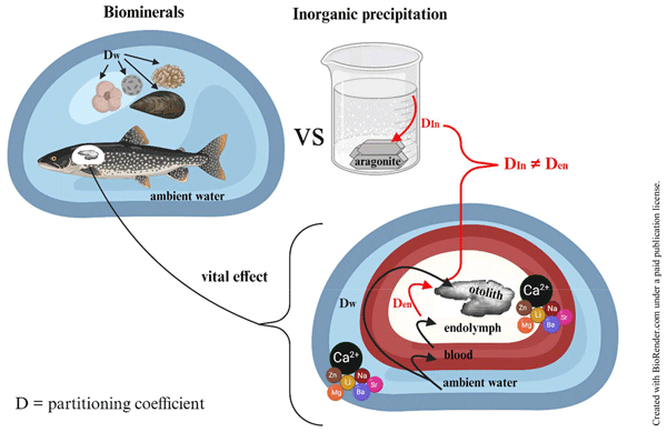
Fish otolith formation is key for understanding the incorporation of elements into biominerals. It is often assumed that the final step of biomineralization consists of inorganic precipitation as the fluid where biominerals form can hardly be sampled. Thanks to fish ear anatomy, this can be overcome with otoliths. By comparing otolith formation and inorganic precipitation, we proved that this assumption is not always true. Our findings could refine models and shed light on biomineralization.
Gerald Langer, Ian Probert, Jeremy R. Young, and Patrizia Ziveri
Biogeosciences, 23, 1795–1808, https://doi.org/10.5194/bg-23-1795-2026, https://doi.org/10.5194/bg-23-1795-2026, 2026
Short summary
Short summary
Coccolithophores are important marine CaCO3 producers and their biominerals, the coccoliths, partly dissolve in the upper water column where dissolution is unexpected. Studying coccolith dissolution in field samples is hampered by a paucity of experimental studies describing dissolution morphologies. Here we fill this gap by experimentally dissolving different coccolithophores and applying our results to field samples.
Falilu O. Adekunbi, Michaël Grelaud, Gerald Langer, Lucian O. Chukwu, Marta Alvarez, Shakirudeen Odunuga, Kai G. Schulz, and Patrizia Ziveri
Biogeosciences, 22, 7865–7880, https://doi.org/10.5194/bg-22-7865-2025, https://doi.org/10.5194/bg-22-7865-2025, 2025
Short summary
Short summary
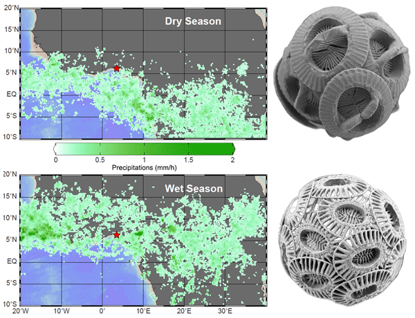
This study is the first to explore seasonal changes in coccolithophores, microscopic algae important for ocean life and the carbon cycle, off the coast of Nigeria. Their abundance and diversity increased during the rainy season, driven by shifts in the Intertropical Convergence Zone. Despite regional differences, these coastal communities show patterns similar to other parts of the world, revealing possible shared environmental pressures.
Stefania Bianco, Manuela Bordiga, Gerald Langer, Patrizia Ziveri, Federica Cerino, Andrea Di Giulio, and Claudia Lupi
Biogeosciences, 22, 1821–1837, https://doi.org/10.5194/bg-22-1821-2025, https://doi.org/10.5194/bg-22-1821-2025, 2025
Short summary
Short summary
This work focuses on the response in culture experiments to increasing CO2 of the coccolithophore species Helicosphaera carteri, a unicellular marine calcifying microalgae. The absence of significant changes in coccolith malformations, along with stable size, shape, and calcification-to-photosynthesis ratio, is indicative of H. carteri low sensitivity to CO2 rise, together with its ability to maintain a stable contribution to the marine rain ratio under future climate changes.
Joost de Vries, Fanny Monteiro, Gerald Langer, Colin Brownlee, and Glen Wheeler
Biogeosciences, 21, 1707–1727, https://doi.org/10.5194/bg-21-1707-2024, https://doi.org/10.5194/bg-21-1707-2024, 2024
Short summary
Short summary
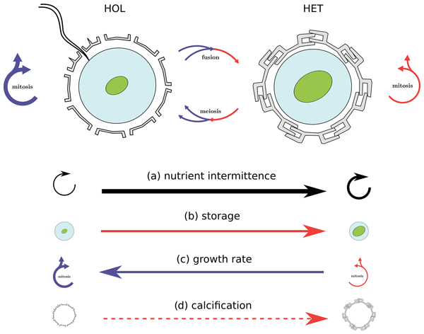
Calcifying phytoplankton (coccolithophores) utilize a life cycle in which they can grow and divide into two different phases. These two phases (HET and HOL) vary in terms of their physiology and distributions, with many unknowns about what the key differences are. Using a combination of lab experiments and model simulations, we find that nutrient storage is a critical difference between the two phases and that this difference allows them to inhabit different nitrogen input regimes.
Cited articles
Balch, W. M., Holligan, P. M., Ackleson, S. G., and Voss, K. J.: Biological and optical properties of mesoscale coccolithophore blooms in the Gulf of Maine. Limnol. Oceanogr., 36, 629–43, 1991.
Baumann, G. F., Isenberg, H. D., and Gennaro, J. J.: The Inverse Relationship Between Nutrient Nitrogen Concentration and Coccolith Calcification in Cultures of the Coccolithophorid Hymenomonas sp., J. Protozool., 25, 253–6, 1978.
Benner, I.: The utilization of organic nutrients in marine phytoplankton with emphasis on coccolithophores, Universität Bremen, Dissertation, 2008.
Bown, P. and Pearson, P.: Calcareous plankton evolution and the Paleocene/Eocene thermal maximum event: New evidence from Tanzania, Mar. Micropaleontol., 71, 60–70, 2009.
Boyd, C. M. and Gradmann, D.: Impact of osmolytes on buoyancy of marine phytoplankton, Mar. Biol., 141, 605–18, 2002.
Buitenhuis, E., van Bleijswijk, J., Bakker, D., and Veldhuis, M.: Trends in inorganic and organic carbon in a bloom of Emiliania huxleyi in the North Sea, Mar. Ecol. Prog. Ser., 143, 271–82, 1996.
Curey, H., Lowenstam, H. A., Epstein, S., and McKinney, C. R.: Measurement of Paleotemperatures and Temperatures of the Upper Cretaceous of England, Denmark, and the Southeastern United States, Geol. Soc. Am. Bull., 62, 399–416, 1951.
De Winter, M. D. A., Schneijdenberg, C. T. W. M., Lebbink, M. N., Lich, B., Verkleij, A. J., Drury, M. R., and Humbel, B. M.: Tomography of insulating biological and geological materials using focused ion beam (FIB) sectioning and low-kV BSE imaging, J. Microsc., 233, 372–83, 2009.
Gerhard, F. and Wefer, G.: Use of Proxies in Paleoceanography. Examples from the South Atlantic. Berlin, Heidelberg, Springer-Verlag, 1999.
Giannuzzi, L. A., Phifer, D., Giannuzzi, N. J., and Capuano, M. J.: Two-dimensional and 3-dimensional analysis of bone/dental implant interfaces with the use of focused ion beam and electron microscopy, J. Oral. Maxillofac. Surg., 65, 737–47, 2007.
Gibbs, S. J., Poulton, A. J., Bown, P. R., Daniels, C. J., Hopkins, J., Young, J. R., Jones, H. L., Thiemann, G. J., O'Dea, S. A., and Newsam, C.: Species-specific growth response of coccolithophores to Palaeocene–Eocene environmental change, Nat. Geosci., 6, 218–22, 2013.
Goodhew, P. J., Humphreys, J. F., and Beanland, R.: Electron Microscopy and Analysis, 3rd Edn., London, New York, Taylor and Francis, 2001.
Grandfield, K. and Engqvist, H.: Focused Ion Beam in the Study of Biomaterials and Biological Matter, Adv. Mater Sci. Eng., 1–6, 2012.
Guillard, R. R. L. and Ryther, J. H.: Studies of marine planktonic diatoms, I, Cyclotella nanna (Hustedt) and Detonula convervacea (Cleve), Can. J. Microbiol., 8, 229–39, 1962.
Hamilton, E. L.: Upper Cretaceous, Tertiary, and Recent planktonic Foraminifera from mid-Pacific flat-topped seamounts, J. Paleontol., 27, 204–37, 1953.
Henriksen, K., Stipp, S. L. S., and Young, J. R.: Biological control on calcite crystallization: AFM investigation of coccolith polysaccharide function, Am. Mineral., 89, 1709–16, 2004.
Holligan, P. M., Fernández, E., Aiken, J., Balch, W. M., Boyd, P., Burkill, P. H., Jones, H. L., Thiemann, G. J., O'Dea, S. A., and Newsam, C.: A biogeochemical study of the coccolithophore, Emiliania huxleyi, in the North Atlantic, Global Biogeochem. Cy., 7, 879–900, 1993.
Holzapfel, C., Schäf, W., Marx, M., Vehoff, H., and Mücklich, F.: Interaction of cracks with precipitates and grain boundaries: Understanding crack growth mechanisms through focused ion beam tomography, Scr. Mater., 56, 697–700, 2007.
Inkson, B. J., Mulvihill, M., and Möbus, G.: 3-D determination of grain shape in a FeAl-based nanocomposite by 3D FIB tomography, Scr. Mater., 45, 753–8, 2001.
Kato, M., Ito, T., Aoyama, Y., Sawa, K., Kaneko, T., Kawase, N., and Jinnai, H.: Three-dimensional structural analysis of a block copolymer by Scanning Electron Microscopy combined with a Focused Ion Beam, J. Polym. Sci. Part. B, 45, 677–83, 2007.
Kelm, K., Goetz, A., Sehrbrock, A., Irsen, S., Hoffmann, R., Schmahl, W. W., and Griesshaber, E.: Mosaic Structure in the Spines of Holopneustes porossisimus, Zeitschrift Für Krist, Cryst. Mater., 227, 758–65, 2012.
Kubis, A. J., Shiflet, G. J., Dunn, D. N., and Hull, R.: Focused Ion-Beam Tomography, Metall Mater. Trans. A, 35, 1935–43, 2004.
Langer, G., Geisen, M., Baumann, K.-H., Kläs, J., Riebesell, U., Thoms, S., and Young, J. R.: Species-specific responses of calcifying algae to changing seawater carbonate chemistry, Geochemistry, Geophys. Geosystems., 7, 1–12, 2006.
Langer, G., Nehrke, G., Probert, I., Ly, J., and Ziveri, P.: Strain-specific responses of Emiliania huxleyi to changing seawater carbonate chemistry, Biogeosciences, 6, 2637–2646, https://doi.org/10.5194/bg-6-2637-2009, 2009.
Langer, G., Probert, I., Nehrke, G., and Ziveri, P.: The morphological response of Emiliania huxleyi to seawater carbonate chemistry changes: an inter-strain comparison, J. Nannoplankton Res., 32, 29–-34, 2011.
Langer, G., Oetjen, K., and Brenneis, T.: Calcification of Calcidiscus leptoporus under nitrogen and phosphorus limitation, J. Exp. Mar. Biol. Ecol., 413, 131–7, 2012.
Langer, G., Oetjen, K., and Brenneis, T.: On culture artefacts in coccolith morphology, Helgol. Mar. Res., 67, 359–69, 2013a.
Langer, G., Oetjen, K., and Brenneis, T.: Coccolithophores do not increase particulate carbon production under nutrient limitation: A case study using Emiliania huxleyi (PML B92/11), J. Exp. Mar. Biol. Ecol., 443, 155–61, 2013b.
Leser, V., Drobne, D., Pipan, Z., Milani, M., and Tatti, F.: Comparison of different preparation methods of biological samples for FIB milling and SEM investigation, J. Microsc., 233, 309–19, 2009.
Menden-Deuer, S. and Lessard, E. J.: Carbon to volume relationships for dinoflagellates, diatoms, and other protist plankton, Limnol. Oceanogr., 45, 569–79, 2000.
Nalla, R. K., Porter, A. E., Daraio, C., Minor, A. M., Radmilovic, V., Stach, E. A., Tomsia, A. P., and Ritchie, R. O.: Ultrastructural examination of dentin using focused ion-beam cross-sectioning and transmission electron microscopy, Micron., 36, 672–80, 2005.
Paasche, E.: Roles of nitrogen and phosphorus in coccolith formation in Emiliania huxleyi (Prymnesiophyceae), Eur. J. Phycol., 33, 33–42, 1998.
Paasche, E.: A review of the coccolithophorid Emiliania huxleyi (Prymnesiophyceae), with particular reference to growth, coccolith formation, and calcification-photosynthesis interactions, Phycologia, 40, 503–29, 2002.
Phaneuf, M. W.: Applications of focused ion beam microscopy to materials science specimens, Micron., 30, 277–88, 1999.
Ridgwell, A., Schmidt, D. N., Turley, C., Brownlee, C., Maldonado, M. T., Tortell, P., and Young, J. R.: From laboratory manipulations to Earth system models: scaling calcification impacts of ocean acidification, Biogeosciences, 6, 2611–2623, https://doi.org/10.5194/bg-6-2611-2009, 2009.
Schmahl, W. W., Griesshaber, E., Merkel, C., Kelm, K., Deuschle, J., Neuser, R. D., ., Goetz, A. J., Sehrbrock, A., and Mader, W.: Hierarchical fibre composite structure and micromechanical properties of phosphatic and calcitic brachiopod shell biomaterials – an overview, Miner. Mag., 72, 541–62, 2008.
Srot, V., Bussmann, B., Salzberger, U., Koch, C. T., and van Aken, P. A.: Linking Microstructure and Nanochemistry in Human Dental Tissues, Microsc. Microanal., 18, 509–23, 2012.
Stoll, H. M. and Schrag, D. P.: Coccolith Sr/Ca as a new indicator of coccolithophorid calcification and growth rate. Geochemistry, Geophys. Geosystems., 1, 1–29, 2000.
Thierstein, H. R. and Young, J. R.: Coccolithophores: From Molecular Processes to Global Impact, Berlin Heidelberg New York, Springer-Verlag, 2004.
Uchic, M. D., Groeber, M. A., Dimiduk, D. M., and Simmons, J. P.: 3-D microstructural characterization of nickel superalloys via serial-sectioning using a dual beam FIB-SEM, Scr. Mater, 55, 23–8, 2006.
Williams, R. E. A., Uchic, M., Dimiduk, D., and Fraser, H. L.: Three-Dimensional Reconstruction of Alpha Laths in Alpha/beta Titanium Alloys by Serial Sectioning with a FEI NOVA, Microsc. Microanal., 11, 836–7, 2005.
Young, J. R.: Functions of coccoliths in: Coccolithophores, edited by: Winter, A. and Siesser, G. W., Cambridge, University Press, 63–82, 1994.
Young, J. R. and Henriksen, K.: Biomineralization Within Vesicles: The Calcite of Coccoliths, Rev. Min. Geochem., 54, 189–215, 2003.
Young, J. R. and Ziveri, P.: Calculation of coccolith volume and it use in calibration of carbonate flux estimates, Deep-Sea Res. Pt. II, 47, 1679–700, 2000.
Young, J. R., Didymus, J. M., Bown, P. R., Prins, B., and Mann, S.: Crystal assembly and phylogenetic evolution in heterococcoliths, Nature, 356, 516–8, 1992.
Young, J. R., Davis, S. A., Bown, P. R., and Mann, S.: Coccolith ultrastructure and biomineralisation, J. Struct. Biol., 126, 195–215, 1999.
Young, R. J., Dingle, T., Robinsonts, K., and Pugh, P. J. A.: An application of scanned focused ion beam milling to studies on the internal morphology of small arthropods, J. Microsc., 172, 81–8, 1993.

Altmetrics
Final-revised paper
Preprint